45 draw and label a compound microscope
UD Virtual Compound Microscope - University of Delaware ©University of Delaware. This work is licensed under a Creative Commons Attribution-NonCommercial-NoDerivs 2.5 License.Creative Commons Attribution-NonCommercial-NoDerivs 2.5 License. Microscopy Pre-lab Activities - University of Delaware Microscope controls: turn knobs (click and hold on upper or lower portion of knob) throw switches (click and drag) turn dials (click and drag) move levers (click and drag) changes lenses (click and drag on objective housing) select a specimen (click on a slide) adjust oculars (in "through" view, with the light on, click and drag to move oculars closer or further apart) Designed and …
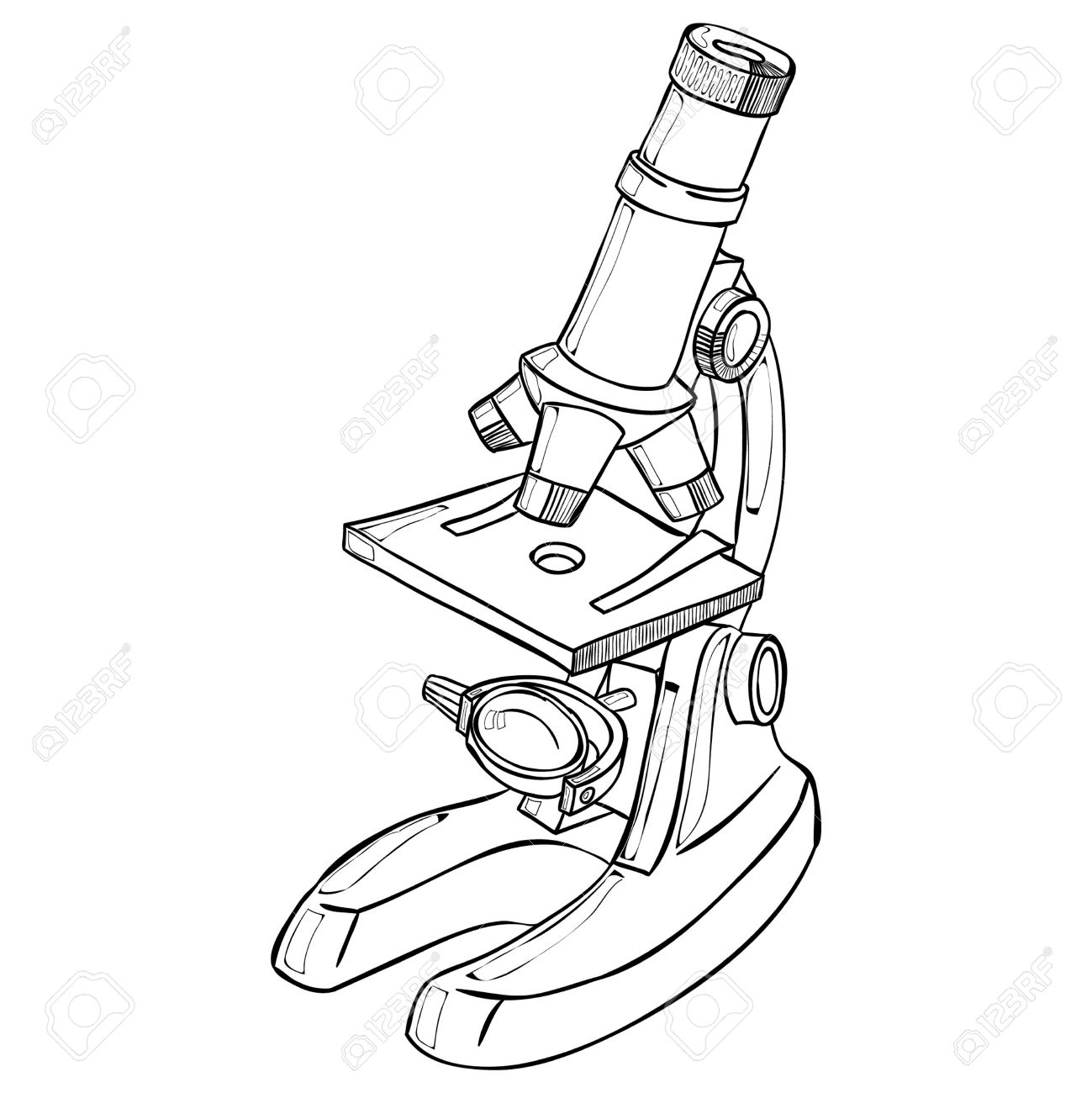
how to draw microscope (compound) - YouTube drawing microscope. Thank you watching more videos.please subscribe my channel

Draw and label a compound microscope
ACTIVITY-1-THE-COMPOUND-MICROSCOPE.pdf - Course Hero ACTIVITY-1-THE-COMPOUND-MICROSCOPE.pdf - Name : Rating : Course : Date : ACTIVITY 1 The Compound Microscope General Instruction: Draw and label a. ACTIVITY-1-THE-COMPOUND-MICROSCOPE.pdf - Name : Rating :... School Cotabato City State Polytechnic College; Course Title Science 123; Uploaded By MateField6213. II - National Council of Educational Research and Training monocot plant (such as lily, maize, grass), compound microscope, slide, cover slip, needle, brush, a piece of blotting paper, and a razor blade. E x p e r i m e n t 23 UNIT II The World of the Living Laboratory Manual Science 94 PROCEDURE 1. Remove a peel from the lower surface of a dicot leaf. This can be easily done by folding or tearing the leaf and pulling out the thin … (b) Why both objective and eyepiece of a compound microscope must have ... (a) Draw the labelled ray diagram for the formation of image by a compound microscope. Derive an expression for its total magnification (or magnifying power), when the final image is formed at the near point. (b) Why both objective and eyepiece of a compound microscope must have short focal lengths?
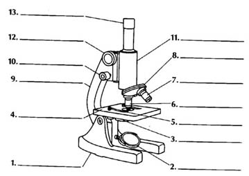
Draw and label a compound microscope. The Compound Microscope.docx - The Compound Microscope 1. Draw and ... Draw and label a compound microscope. Parts Functions Parts of a Compound Microscope 2. Enumerate all the parts of the microscope and give their corresponding functions. Tabulate your answer. a. Objective Lenses -forms the inverted image of the specimen and gives the initial magnification. -used for visualization of specimen. c. What are the Basics of Biochemical Techniques? 28.04.2022 · The transmission electron microscope (TEM) works much like a light microscope, transmitting a beam of electrons through a thin specimen and then focusing the electrons to form an image on a screen or on film. This is the most common form of an electron microscope and has the best resolution. The scanning electron microscope (SEM) scans a fine beam of … Parts of a microscope with functions and labeled diagram Q. List down the 18 parts of a Microscope. 1. Ocular Lens (Eye Piece) 2. Diopter Adjustment 3. Head 4. Nose Piece 5. Objective Lens 6. Arm (Carrying Handle) 7. Mechanical Stage 8. Stage Clip 9. Aperture 10. Diaphragm 11. Condenser 12. Coarse Adjustment 13. Fine Adjustment 14. Illuminator (Light Source) 15. Stage Controls 16. Base 17. Labelled Diagram of Compound Microscope - Biology Discussion The below mentioned article provides a labelled diagram of compound microscope. Part # 1. The Stand: The stand is made up of a heavy foot which carries a curved inclinable limb or arm bearing the body tube. The foot is generally horse shoe-shaped structure (Fig. 2) which rests on table top or any other surface on which the microscope in kept.
PRACTICAL BOOKLET - BIOLOGY4ISC Aim: To Prepare Stained Temporary Mount of Onion Peel Materials Required: Glass slide, cover slip, watch glasses, dropper, forceps, mounting needle, brush, blotting paper, compound microscope, knife/scalpel, onion, glycerine, safranin solution, distilled water. Procedure. Pour some distilled water into a watch glass. Take a piece of onion and with the help of a forcep … Draw a labelled ray diagram of a compound microscope and explain its ... In this case, the objective lens O of the compound microscope forms a real, inverted and enlarged image A'B' of the object. Now A'B' acts as an object for the eyepiece E, whose position is adjusted so that A'B' lies between optical centre C2 and the focus fe' of eyepiece. Now the eyepiece forms a final virtual, inverted and highly ... Compound Microscope: Definition, Diagram, Parts, Uses, Working ... - BYJUS A compound microscope is defined as A microscope with a high resolution and uses two sets of lenses providing a 2-dimensional image of the sample. The term compound refers to the usage of more than one lens in the microscope. Also, the compound microscope is one of the types of optical microscopes. Parts of the Microscope with Labeling (also Free Printouts) 5. Knobs (fine and coarse) By adjusting the knob, you can adjust the focus of the microscope. The majority of the microscope models today have the knobs mounted on the same part of the device. Image 5: The circled parts of the microscope are the fine and coarse adjustment knobs. Picture Source: bp.blogspot.com.
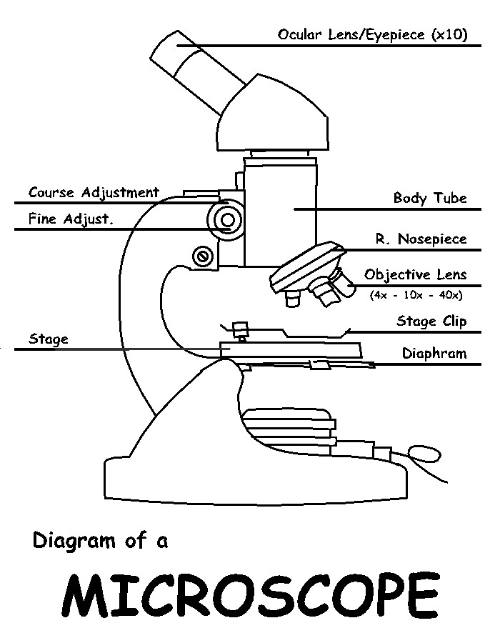
Parts of the Compound Microscope - HCC Learning Web Parts of the Compound Microscope Use Figure 2 as a guide to locate the major parts of the compound microscope. a. Base: The bottom, flat part that supports the microscope. b. Arm: The straight or curved vertical part that connects the base to the upper portion. c. Body Tube: Extends from the arm and contains the ocular lens and the rotating How to draw compound of Microscope easily - step by step I will show you " How to draw compound of microscope easily - step by step "Please watch carefully and try this okay.Thanks for watching.....#microscopedrawi... anyflip.com › jiefu › wljfChemistry Form 4 KSSM Text Book - Flip eBook Pages 1-50 | AnyFlip Oct 15, 2021 · (a) Copy Figure 1 and label the melting point of lauric acid, C12H24O2 on the diagram. Time (min) Figure 1 (b) Draw the arrangement of particles in lauric acid, C12H24O2 between R and S. (c) The melting point of lauric acid, C12H24O2 is 43 °C. Suggest a suitable method of heating lauric acid, C12H24O2. (d) Draw a labelled diagram to show the ... (i) Draw a neat labelled ray diagram of a compound microscope. Explain ... 65.2k views asked May 15, 2018 in Physics by paayal (148k points) (i) Draw a neat labelled ray diagram of a compound microscope. Explain briefly its working. (ii) Why must both the objective and the eye-piece of a compound microscope have short focal lengths? cbse class-12 1 Answer 0 votes answered May 15, 2018 by sanjaydas (89.4k points)
34 Label Compound Light Microscope - Labels Database 2020
Compound Microscope - Diagram (Parts labelled), Principle and Uses What are the 13 parts of a microscope? 1. Eyepiece 2. Eyepiece Tube 3. Objective Lens 4. Stage 5. Stage Clips 6. Nosepiece 7. Fine and Coarse Focus knobs 8. Illuminator 9. Aperture 10. Iris Diaphragm 11. Condenser 12. Condenser Focus Knob 13. The Rack stop Q 5. What are the 11 parts of a compound microscope?

Cartoon Simple Microscope Drawing - Micropedia
Cell Types Gizmo Worksheet - StuDocu Gizmo Warm-up In the Cell Types Gizmo, you will use a light microscope to compare and contrast different samples. On the LANDSCAPE tab, click on the Elodea leaf. (Turn on Show all samples if you can’t find it.) Switch to the MICROSCOPE tab to observe the sample as it would appear under the microscope. By default, this microscope is using 40x ...
Free Microscope Drawing, Download Free Microscope Drawing png images ...
Label the microscope — Science Learning Hub Use this with the Microscope parts activity to help students identify and label the main parts of a microscope and then describe their functions. Drag and drop the text labels onto the microscope diagram. If you want to redo an answer, click on the box and the answer will go back to the top so you can move it to another box.

Microscope Labeled Diagram - Micropedia
Compound Microscope Parts, Functions, and Labeled Diagram Compound Microscope Definitions for Labels. Eyepiece (ocular lens) with or without Pointer: The part that is looked through at the top of the compound microscope. Eyepieces typically have a magnification between 5x & 30x. Monocular or Binocular Head: Structural support that holds & connects the eyepieces to the objective lenses.

A Well Labelled Diagram Of A Light Microscope - Top Label Maker
› LabManuals › biol123STAINING OF BACTERIAL CELLS Objective Introduction 7. Flame the loop again until the wire glows. Cool the loop and draw a sample from another plate and place in the second circle. With circular motion of the loop, dissolve the cells in the water. 8. Repeat the above steps for the second glass slide, making sure you label the slides so that you know which organisms are on each slide. 9.
Microscope Drawing Template at GetDrawings | Free download
Microscope Parts, Function, & Labeled Diagram - slidingmotion Objective lenses. Objective lenses are the most important part of the microscope. Its purpose is to visualize the specimen. There are 3-4 types of different objective lenses in any microscope. It has a magnification power of 4X to 100 X. 4X objective lens is the shortest lens while the 100X lens is the longest in terms of visualization.

20+ Fantastic Ideas Drawing Diagram Microscope Images - Cine Regard
Microscope Parts and Functions First, the purpose of a microscope is to magnify a small object or to magnify the fine details of a larger object in order to examine minute specimens that cannot be seen by the naked eye. Here are the important compound microscope parts... Eyepiece: The lens the viewer looks through to see the specimen.
Free Microscope Drawing, Download Free Microscope Drawing png images ...
biology4isc.weebly.com › practical-bookletPRACTICAL BOOKLET - BIOLOGY4ISC The first microscope was constructed by Anton Van Leeuwenhoek (1632-1723). This, microscope consisted of a single biconvex lens fitted in a small window of a “board” and the object was viewed through it. This was a simple microscope. After this compound microscope, were developed using combinations of two lenses.

Compound Microscope Diagram Class 11 - Micropedia
Compound Microscope - Types, Parts, Diagram, Functions and Uses It comes with a wide body and base. Its distinct parts include a condenser, illumination, focus lock, mechanical stage, and a revolving nosepiece which can hold up to five objectives. It usually has a binocular head, which makes long-term observation easy. Image 22: An example of a research compound microscope.
Post a Comment for "45 draw and label a compound microscope"