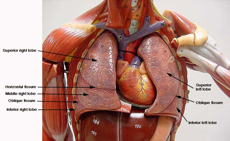
38 label the structures of the thoracic cavity.
Thoracic Bones Anatomy & Structure | What is the Rib Cage? - Study.com Thoracic Cage Bones. The thoracic cage encloses the thorax and the organs within it. It is located at the center of the thorax and is commonly referred to as the rib cage. The thoracic cage bones ... Respiratory System Questions and Answers | Homework.Study.com Place the following structures in the order in which air will reach them when breathing in: (a) bronchus, (b) trachea, (c) nasal (d) cavity, (e) alveolus. View Answer Describe what happens to oxygen and carbon dioxide as blood travels through the circulatory system that starts in the left ventricle and finishes in the left atrium.
thoracic cavity | Description, Anatomy, & Physiology | Britannica thoracic cavity, also called chest cavity, the second largest hollow space of the body. It is enclosed by the ribs, the vertebral column, and the sternum, or breastbone, and is separated from the abdominal cavity (the body's largest hollow space) by a muscular and membranous partition, the diaphragm.

Label the structures of the thoracic cavity.
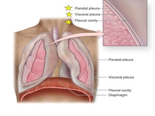
Thoracic cavity - Wikipedia Structures within the thoracic cavity include: structures of the cardiovascular system, including the heart and great vessels, which include the thoracic aorta, the pulmonary artery and all its branches, the superior and inferior vena cava, the pulmonary veins, and the azygos vein Body Cavities and Membranes: Labeled Diagram, Definitions - EZmed Body Cavities Labeled Diagram: There are 2 main cavities in the body, the dorsal cavity and the ventral cavity. We know from the anatomical directional terms lecture that anterior means front or toward the front of the body, and posterior means back or toward the back of the body. Anatomy Chapter 1: Labeling Thoracic Cavity Diagram | Quizlet The cavities surrounding each lung parietal pleura The aspect of the pleura that does not touch the surface of the lung visceral pleura The aspect of the pleura that covers the external surface of the lung The thoracic cavity can be subdivided into... 1. mediastinum 2. left and right pleural cavities 3. pericardial cavity
Label the structures of the thoracic cavity.. Pathology Outlines - Myoepithelioma 08.09.2021 · Epithelial component should be less than 5% (some consider even focal epithelial differentiation sufficient to label the tumor as pleomorphic adenoma) (J Oral Maxillofac Pathol 2013;17:257) Monomorphic histology and rare or absent ductal structures in myoepithelioma differentiate it from pleomorphic adenoma Thoracic Examination - Physiopedia The Thoracic Spine has a complex and often overlooked role within the body. It is a key area of load transfer between the upper and lower body and for rotational movement within the body. Should be assessed and treated as a functional unit including not only the spine but the rib cage.; The thoracic region provides a site for muscle and connective tissue attachments from the … AP 2 (part 3) Flashcards | Quizlet Correctly label the following anatomical features of the lymph node. Label the structures of the spleen. Label the structures of the spleen. Match the lymphatic trunk with the major body region that it drains. Intestinal trunks: Drain most abdominal structures. Lumbar trunks: Drain lower limbs and pelvic organs. Jugular trunks: Label the thoracic cavities.docx - Label the cavities... In the figure above - locate the thoracic cavity. Labelthe structure that separates the thoracic cavity from the abdominopelvic cavity Notice the 4 colors of the thoracic cavity. There are two purple cavities within the thoracic cavity. Labelthem. Identifythe two / three primary structures that lie within the purple cavity. Notice the green cavity.
Label Thoraic Cavity 2.png - l View site information l... View Homework Help - Label Thoraic Cavity 2.png from BIO 141 at Northern Virginia Community College. l View site information l Label the structures of the thoracic Study Resources Main Menu Organs in the Thoracic Cavity - Bodytomy The thoracic cavity is lined by a serous membrane that exudes a thin fluid (serum). The chest membrane, also known as parietal pleura, extends further to cover the lungs. This part of the membrane is known as the visceral pleura. The part of the membrane that covers the heart, esophagus, and the great vessels is known as mediastinal pleura. 681 Thoracic cavity Images, Stock Photos & Vectors - Shutterstock Find Thoracic cavity stock images in HD and millions of other royalty-free stock photos, illustrations and vectors in the Shutterstock collection. Thousands of new, high-quality pictures added every day. Answered: since red blood cells lack… | bartleby Define and describe how the thoracic cavity changes in size and shape during respiration. A: We can say that In human physiology, the movement of the oxygen from the environment to the cells… question_answer. Q: Some muscles that control the tongue and larynx are attached to the a. maxillae. b. cervical… A: We can say that The muscular system is defined as the organ …
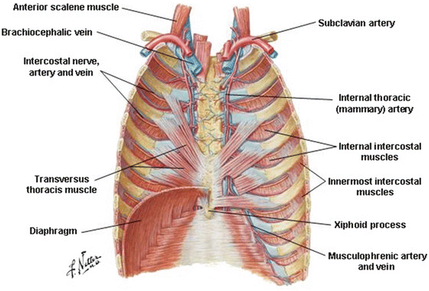
Lab 8: Dissection: Chest Wall, Overview of Thoracic Cavity 3 Open the pleural sacs and define the pleural cavity, parietal pleura, and visceral pleura. 4 Remove the right lung. 5 Strip pleura from right side and clean structures of the posterior wall. 6 Clean and demonstrate the structures in the superior mediastinum. 7 Review the blood supply and venous drainage of the thoracic wall. 8 Identify the ... Thorax: Anatomy, wall, cavity, organs & neurovasculature | Kenhub It is made up of the sternum, twelve pairs of ribs, twelve thoracic vertebrae, and interconnecting joints. The main thoracic joints include the intervertebral discs, costovertebral, sternocostal, sternoclavicular, costochondral, and interchondral joints. Running between every two adjacent ribs are anatomical spaces called intercostal spaces. Ch. 15 Cardiovascular Flashcards | Quizlet During inspiration, the pressure in the thoracic cavity is _____ the pressure in the abdominal cavity. less than. Therefore, during inspiration, the difference in pressure causes blood to flow from the _____ cavity toward the _____ cavity. abdominal; thoracic. Which of the following gases does endothelium release? Nitric oxide. Label the components of the walls of the artery … A&P 139 Chapter 19 Flashcards | Quizlet Label the structures of the nasal cavity. A mother and two young children are found passed out in their apartment, where a space heater is on. Emergency medical technicians suspect carbon monoxide poisoning, so they give the patients . highly concentrated oxygen and some carbon dioxide. laryngeal cartilages. Label the photomicrogram of the lung. Label the photomicrogram …
Anatomy of the Thoracic Wall, Pulmonary Cavities, and ...
Thoracic Cavity - Anatomy | Organs | Functions | 8 Types of Cavities Thoracic Cavity Right and left serous membrane cavities (contain right and left lungs) Mediastinum: Higher portion stuffed with blood vessels, trachea, esophagus, and thymus. The lower portion contains pericardial space (the heart is found at intervals the serosa cavity) 3. Serous Membranes Line of body cavities and canopy organs. Consist of

Summary Sheet 1 Questions - Human Anatomy and Physiology ...
Ch. 19 Circulatory System- heart Flashcards | Quizlet The first and last structures are given. Right atrium 1. tricuspid valve 2. right ventricle 3. pulmonary valve 4. pulmonary trunk 5. pulmonary artery 6. lungs 7. pulmonary vein 8. left atrium Left atrioventricular valve Recommended textbook solutions Essentials of Human Anatomy and Physiology 8th Edition Elaine N. Marieb 648 solutions
Torsos
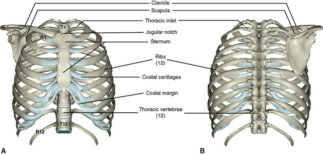
Anatomy: Thoracic Cavity - RnCeus.com The thoracic cavity is made up of 12 pairs of ribs that connect in the posterior thorax to the vertebral bodies of the spinal column. In the anterior thorax, the first 7 pairs of ribs are attached to the sternum or breastbone by cartilage. The lower 5 ribs do not attach to the sternum. The 8th, 9th, and 10th ribs are attached to each other by ...

Unit 1 Lab Homework Flashcards | Quizlet
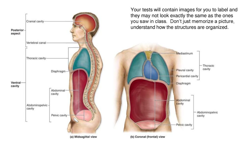
Anatomical Body Planes | Science Trends Examples of these anatomical terms used to label body structures include the axial skeleton, the median cerebral artery, the posterior and anterior pituitary, and the inferior and superior vena cava. Prefixes and suffixes are also used to indicate the position of anatomical structures within the body. The parathyroid glands have the prefix Para attached to them, referencing the fact …

Thoracic wall and breast (Illustrations) - e-Anatomy
lining of the thoracic cavity - Microsoft lining of the thoracic cavity. Pig fetal stomach system lining digestive inner dissection appearance describe. Serous membranes membrane body anatomy cavity cavities ventral organs human serosa heart pleural thoracic. Pig fetal gallbladder digestive describe system liver dissection location lobes lab does. Body cavities and membranes : Anatomy ...
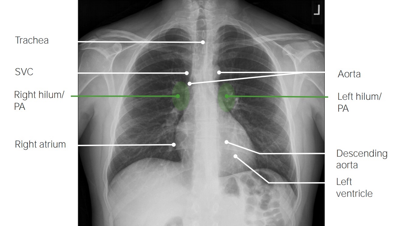
Imaging of the Lungs and Pleura | Concise Medical Knowledge
The Thoracic Cavity - Human Anatomy The Thoracic Cavity. The heart and lungs are situated in the thorax, the walls of which afford them protection. The heart lies between the two lungs, and is enclosed within a fibrous bag, the pericardium, while each lung is invested by a serous membrane, the pleura. The skeleton of the thorax, and the shape and boundaries of the cavity, have ...

Professional Medical Anatomy of Human Organ System Trunk Thoracic Cavity Structure Model of The Internal Organs
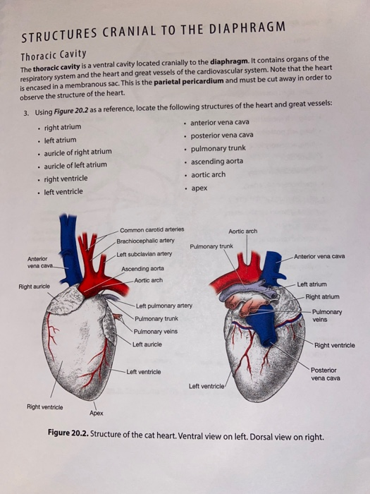
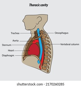
Pre-Lab Exercise 16-2 Anatomy of the Thoracic Cavity Label and color ... Pre-Lab Exercise 16-2 Anatomy of the Thoracic Cavity Label and color the structures in the thoracic cavity in Fig text and Exercise 16-1 in this unit for reference 16with the terms from Excise 16-161931. Use your FIGURE 16.1 The thoracic cavity Cardiovascular System-Part1: The Heart I UNIT 16 Pre-Lab Exercise 16-3 Anatomy of the Heart Label and color the three views of the heart in Figure 16.2 ...

Human Skeleton System Thoracic Skeleton with Label Design ...
Solved Label the structures of the thoracic cavity. Trachea | Chegg.com Anatomy and Physiology. Anatomy and Physiology questions and answers. Label the structures of the thoracic cavity. Trachea Diaphragm Esophagus. Question: Label the structures of the thoracic cavity. Trachea Diaphragm Esophagus.

Thoracic Cavity Labeling Diagram | Quizlet
2010 ACCF/AHA/AATS/ACR/ASA/SCA/SCAI/SIR/STS/SVM … The second step is to confirm that the dissection flap has a motion independent of surrounding structures and that the apparent flap is contained within the aortic lumen (ie, does not pass through the aortic wall in any view). The third step is to use color-flow Doppler to demonstrate differential flow on the 2 sides of the dissection flap. Often one can visualize 1 or more sites of …

LABEL ORGANS IN THORACIC CAVITY Diagram | Quizlet
Pain | definition of pain by Medical dictionary Pain Definition Pain is an unpleasant feeling that is conveyed to the brain by sensory neurons. The discomfort signals actual or potential injury to the body. However, pain is more than a sensation, or the physical awareness of pain; it also includes perception, the subjective interpretation of the discomfort. Perception gives information on the pain's ...
thoracic cavity | Description, Anatomy, & Physiology | Britannica
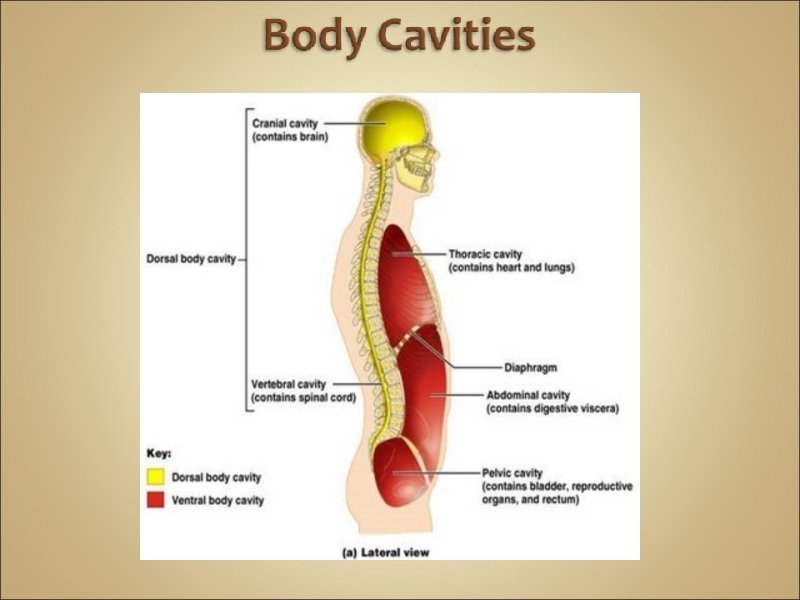
Abdominal Cavity - Definition and Organs | Biology Dictionary Cranial cavity - The space found within a skull, which is occupied by the brain. Rib cage - A bony structure that has 12 pairs of ribs and that houses and protects the lungs and heart. Thoracic cavity - The space in which the rib cage, heart, and lungs are found in vertebrates. Trachea - The tube that carries air between the larynx and ...

APR Lymphatic System Flashcards | Quizlet
Thoracic cage: Anatomy and clinical notes | Kenhub The thoracic cage, also known as the rib cage, is the osteocartilaginous structure that encloses the thorax.It is formed by the 12 thoracic vertebrae, 12 pairs of ribs and associated costal cartilages and the sternum.. The thoracic cage takes the form of a domed bird cage with the horizontal bars formed by ribs and costal cartilages. It is supported by the vertical sternum (anteriorly) and the ...

Pin on School Stuff
Answered: Label the following: Femur * Head *… | bartleby Solution for Label the following: Femur * Head * Neck * Greater trochanter * Lesser trochanter * Medial condyle * Lateral condyle Medial epicondyle * Lateral…
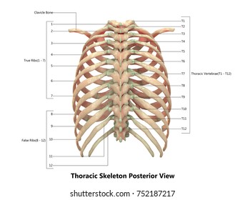
Human Skeleton System Thoracic Skeleton Anatomy Stock ...
Unit 1 Lab Homework Flashcards | Quizlet Terms in this set (13) Label the regions of the body. Label the structures of the thoracic cavity. Label the directional terms based on the arrows. Label the body planes. Label the directional terms based on the directions of the arrows. _____ is towards the front of the body. _____ is farther from the trunk or origin of a structure.

Label Thoraic Cavity 2.png - l View site information l Label ...
Thoracic Cavity - Introduction, Structure, Organs, and FAQs - VEDANTU In the centre of the chest between the lungs is the mediastinum that comprises the organs that are located inside it. Structures within the thoracic cavity include: Oesophagus of the digestive system Thymus gland Vagus nerve and parasympathetic veins. Diaphragm, trachea, bronchi, lungs. The heart The superior and inferior vena cava.
Edited Respi
Chapter 8/10 Flashcards | Quizlet Label the structures of the bone using the hints provided. ... Correctly label the muscles of the thoracic cavity and abdomen. The concave surface of the ulna that wraps around the trochlea of the humerus is the _____ _____. trochlear notch. Recommended textbook solutions.
3: The Thorax | Pocket Dentistry
points Label the structures of the thoracic cavity. Parietal pleura ... Question: Award: 0.76 points Label the structures of the thoracic cavity. Parietal pleura Visceral pleura Pleural cavity Parietal pericardium Visceral perica...
Thorax: Anatomy, wall, cavity, organs & neurovasculature | Kenhub
Thoracic Cavity - Definition & Organs of Chest Cavity - Biology Dictionary The thoracic cavity is actually composed of three spaces each lined with mesothelium, a special film-like tissue that separates vital organs. The pleural cavities surround the lungs, while the pericardial cavity surrounds and protects the heart. These tissues in the thoracic cavity can be seen in the image below.
Anatomy of the Thoracic Wall, Pulmonary Cavities, and ...
Anatomy Chapter 1: Labeling Thoracic Cavity Diagram | Quizlet The cavities surrounding each lung parietal pleura The aspect of the pleura that does not touch the surface of the lung visceral pleura The aspect of the pleura that covers the external surface of the lung The thoracic cavity can be subdivided into... 1. mediastinum 2. left and right pleural cavities 3. pericardial cavity
Introduction to Human Anatomy and Physiology Anatomy the
Body Cavities and Membranes: Labeled Diagram, Definitions - EZmed Body Cavities Labeled Diagram: There are 2 main cavities in the body, the dorsal cavity and the ventral cavity. We know from the anatomical directional terms lecture that anterior means front or toward the front of the body, and posterior means back or toward the back of the body.

Ventral Body Cavity | Subdivisions, Organs, & Diagram - Video ...
Thoracic cavity - Wikipedia Structures within the thoracic cavity include: structures of the cardiovascular system, including the heart and great vessels, which include the thoracic aorta, the pulmonary artery and all its branches, the superior and inferior vena cava, the pulmonary veins, and the azygos vein

Thoracic Images, Illustrations & Vectors (Free) - Bigstock

Label the organs 1. brain 2. Thyroid gland 3. Trachea 5 ...
Thorax: Anatomy, wall, cavity, organs & neurovasculature | Kenhub

Solved Correctly label the following structures related to ...
Thoracic Cavity - Definition & Organs of Chest Cavity ...
Solved Using the figures provided, label the numbered | Chegg.com
Introduction to Human Anatomy and Physiology (Chapter 1 ...
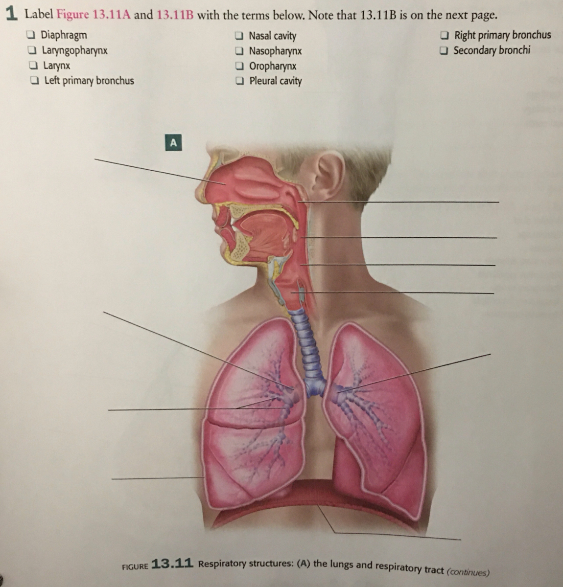
Answered: I Label Figure 13.11A and 13.11B with… | bartleby

A&P - Anatomy & Physiology: The Unity of Form and Function ...
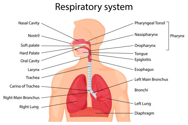
97 Diagram Of The Respiratory System With Labels Drawing ...
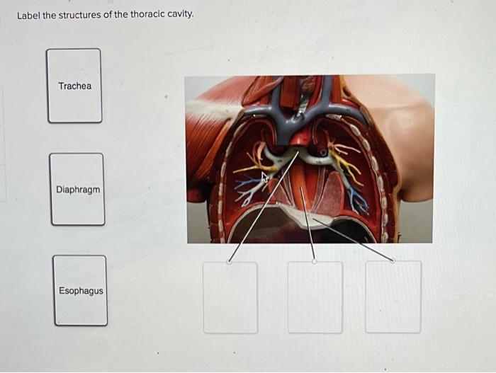
Solved Label the structures of the thoracic cavity. Trachea ...
683 Thoracic cavity Images, Stock Photos & Vectors | Shutterstock
Pleural cavity: Anatomy, location, function | Kenhub

The thoracic cavity | Thoracic cavity, Respiratory system ...
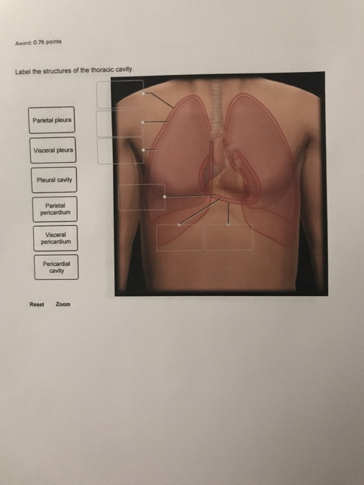
Solved Award: 0.76 points Label the structures of the | Chegg.com
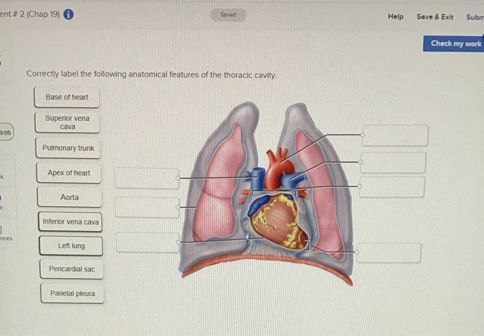
Solved ent # 2 (Chap 19) Help Save & Exit Subm Check my work ...

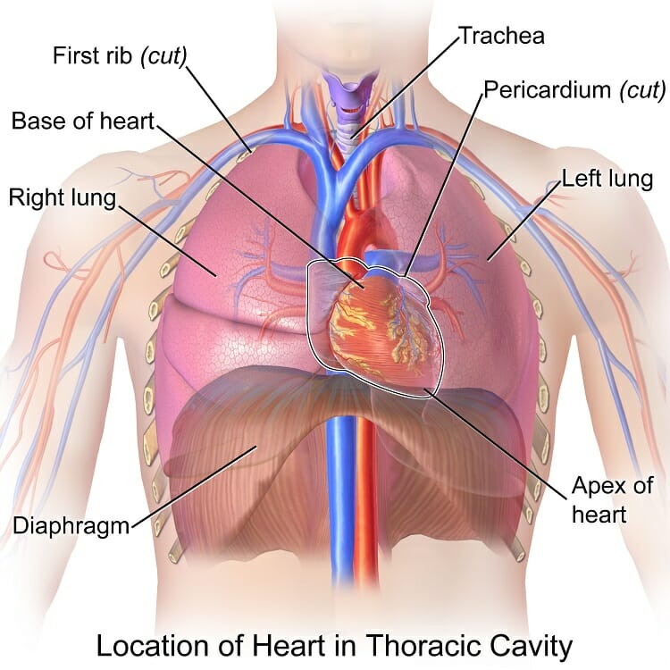
Figure 27.1 Heart in thoracic cavity Diagram | Quizlet
Post a Comment for "38 label the structures of the thoracic cavity."