43 dental x-ray tube head diagram
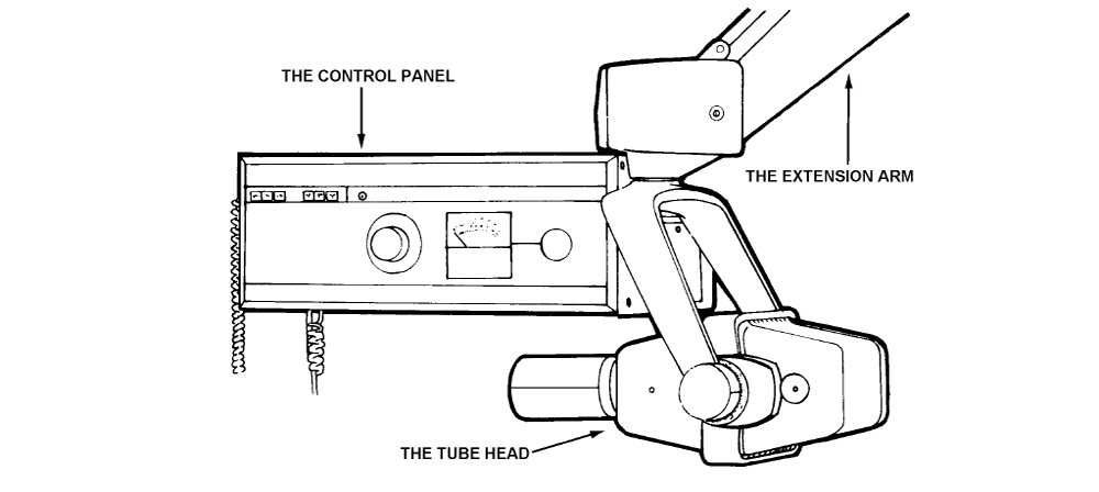
PDF Easy Guide to Dental X-ray Positioning - AAHA Easy Guide to Dental X-ray Positioning . Canine. Tooth Tube Head Angle Comments Maxilla . 110-109 & 210-209 (Molars) 45-55° High and thru the eye ... tube head parallel to the nose crack 103-203 (All incisors) 45-60° Parallel to nose crack . Mandible . 311 -308 & 411 -408 (Molars) Lateral to sensor Don't place the sensor too deep for 311 & ... PDF ENTALALEZ EZ TECH ECH TTIPS IPS - Frank's Hospital Workshop of HDX Intraoral X-ray Installation, Operation and Maintenance Manual, p/n 353002 or page 30 and 31 of HDX Intraoral X-ray Installation, Operation and Maintenance Manual, p/n 353010.) Tube Head Drifting: A plastic bearing in the distal arm functions as a brake for the tube head. To make adjustments, remove distal arm cover and find the set screw
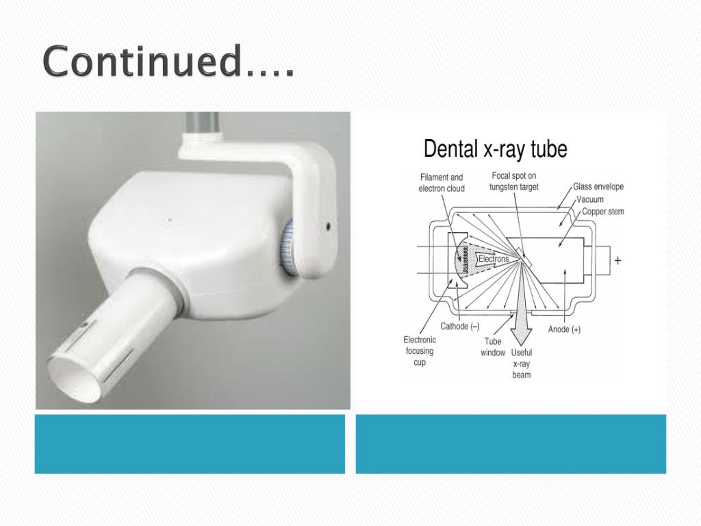
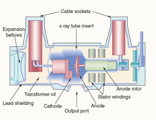
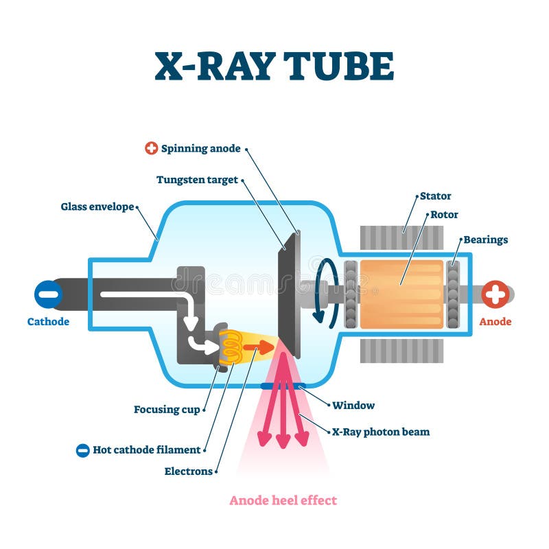
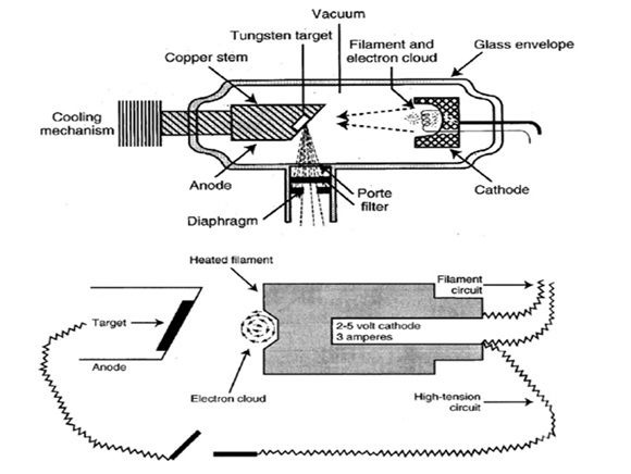
The X-ray Tube | Radiology Key The general-purpose x-ray tube is an electronic vacuum tube that consists of an anode, a cathode, and an induction motor all encased in a glass or metal enclosure (envelope). Figure 5-3 provides a labeled illustration of this design. Recall that the anode is the positive end of the tube and the cathode is the negative end of the tube.
Dental x-ray tube head diagram
Facial trauma - Wikipedia Facial trauma, also called maxillofacial trauma, is any physical trauma to the face.Facial trauma can involve soft tissue injuries such as burns, lacerations and bruises, or fractures of the facial bones such as nasal fractures and fractures of the jaw, as well as trauma such as eye injuries.Symptoms are specific to the type of injury; for example, fractures may involve pain, … Dental X-ray Equipment Parts - Find Local Dentist Near Your Area Dental X-ray Equipment Parts. 25 Aug 2011 Intraoral Receptors, Dental Receptors placed inside the mouth. Tube Head, part of the dental x-ray machine that contains the x-ray tube. Dental X-ray Equipment Parts. 25 Aug 2011 the component part of the dental x-ray machine that allows movement and positioning of the tubehead is termed the, position ... PDF INSTALLATION INSTRUCTIONS - Belmont Equipment x-ray tube, high voltage generator or both. B. Duty cycle A cool down interval of 50 seconds or more must be allowed between each 1 second exposure. (a 25 second cool down must be allowed between each 0.5 second exposure.) This will avoid the accumulation of excess heat and prolong the tube head life. C. Tube head cooling curve 0 0
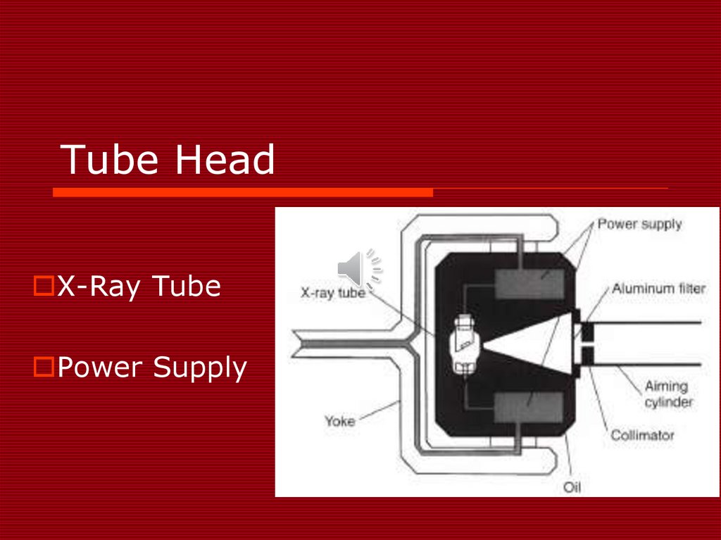
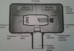
Dental x-ray tube head diagram. RADIOLOGY CH 2 Dental X-ray Tubehead Diagram - Quizlet surrounds the X-ray tube and transformers and is filled with oil. protects the X-ray tube and grounds the high-voltage components. Location. tube head and x-ray tube components Diagram | Quizlet x-ray tube unleaded glass, heart of the X-ray generating system transformer (the power supply) alerts the voltage of incoming electrical current. collimator Forms the size and shape of the X-ray beam aluminum filter disks that filter out long wavelengths PID lead lined cylinder, positions towards patients mouth. cathode - generate electrons Label the Dental X-ray Tubehead (Screencast) - Wisc-Online The tubehead is a sealed, heavy metal housing that contains the x-ray tube that produces dental x-rays. This learning object will provide students with ... PDF DENTAL X-RAY 097 - Belmontdental the x-ray tube, high voltage generator or both. B. Duty cycle A cool down interval of 50 seconds or more must be allowed between each 1 second exposure. (a 25 second cool down must be allowed between each 0.5 second exposure.) This will avoid the accumulation of excess heat and prolong the tube head life. C. Tube head cooling curve 1.
US4157476A - Dental X-ray tube head - Google Patents In a dental x-ray tube head, the x-ray tube is in a casing that supports the tube and shields against stray radiation being projected to the environment through the housing of the tube head. The... X-ray tube - Wikipedia As time goes on, the tube becomes unstable even at lower voltages, and must be replaced. At this point, the tube assembly (also called the "tube head") is removed from the X-ray system, and replaced with a new tube assembly. The old tube assembly is shipped to a company that reloads it with a new X-ray tube. Labelling Dental X-Ray Tube head Diagram - Quizlet Start studying Labelling Dental X-Ray Tube head. Learn vocabulary, terms, and more with flashcards, games, and other study tools. Home | Hamamatsu Photonics The official website of Hamamatsu Corporation whose mission is to advance science and industry through photonic technologies. Our products include optical sensors and components, cameras, light & radiation sources, lasers, and customized solutions.
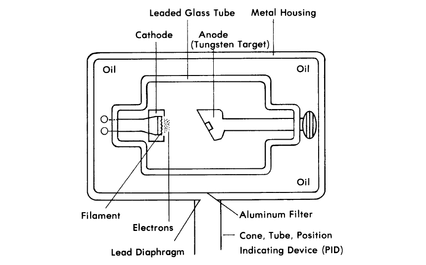
NEA | Additional Licensing Information Dental Radiography (Dentist) Registration as a dentist under Singapore Dental Council. For dentists under Division 2 of the Singapore Dental Council register that did not undergo formal training (i.e. trained through apprenticeship), the dentist will not be permitted to energise an ionising apparatus. Work with Enclosed X-Ray System- Can a Sinus Infection Be Caused by a Tooth? | Oral Answers Had an nfection up by my sinus area that showed up in a dental x-ray about 3 years ago. It, according to my regular dentist, was caused by a bad tooth (upper right molar). This dentist upon seeing my x-rays told me he couldn’t help me, that I had to go to an endodontist for this problem and recommended some. Production of Dental X-Rays - Dental Radiology - YouTube A summary of how an xray is produced in the dental xray tube head.Information taken from:Dental Radiography Principle and Techniques - by Iannucci and Howert... Steps In The Process Of Xray Production - Dental Radiography See figures 1-3, 1-4, and 1-5 for a diagram of the complete procedure. Figure 1-3. Tube head with the filament of the cathode emitting electrons. Figure 1-4. Electrons speeding toward the anode (tungsten target). Leaded Glass Tube Metal Housing Cathode Anode (Tungsten Target) Cathode Anode (Tungsten Target) Aluminum Filter
PPT - DENTAL RADIOGRAPH. PowerPoint Presentation, free ...
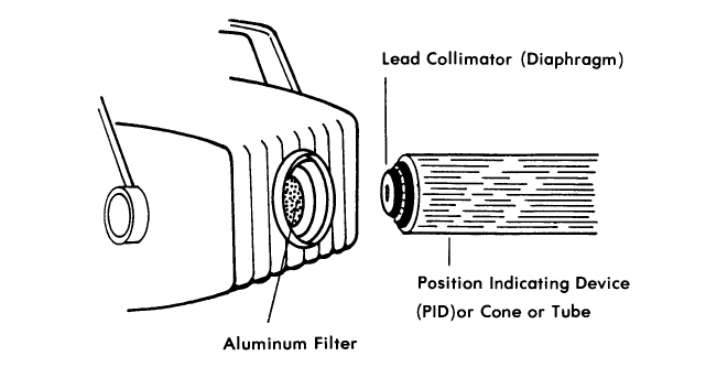
Radiation Protection - Welcome to Dental Radiography The following diagram will identify the location of these two devices (see figure 1-8). Figure 1-8. Tube head assembly: filter, collimator (diaphragm), PID or cone or tube. b. Filter. The aluminum filter or disk is placed in the path of the x-ray beam. Figure 1-8 shows the location of the PID.
X-Ray Machine - презентация онлайн
Safety Code 35: Safety Procedures for the Installation, Use and … However, in systems where an X-ray tube for radiography is also present, the shielding for this X-ray tube must be evaluated independently, as in Section B1.3.2. When equipment include more than one X-ray tube, such as in cardiac systems, the shielding calculation must take into account each X-ray tube independently.

tube head and x-ray tube components Diagram | Quizlet
US4575330A - Apparatus for production of three-dimensional A system for generating three-dimensional objects by creating a cross-sectional pattern of the object to be formed at a selected surface of a fluid medium capable of altering its physical state in response to appropriate synergistic stimulation by impinging radiation, particle bombardment or chemical reaction, successive adjacent laminae, representing corresponding successive …

How Does Your X-Ray Work? – Welcome to R-Tech Dental
Top Dental Digital Radiography Mistakes - DentalSensors.com Then make sure your x-ray head tube is flush against the ring. There should be less than an inch gap between the end of the x-ray head tube and the patients skin. X-ray head generators are a lot like a shot gun. The farther you are away from your target or in your case a dental sensor. The less you are going to hit that target.

Dentstudy - 👉DENTAL X-RAY TUBE HEAD ⭐ X-rays are produced ...
X-ray tube - Wikipedia History. X-ray tubes evolved from experimental Crookes tubes with which X-rays were first discovered on November 8, 1895, by the German physicist Wilhelm Conrad Röntgen.These first generation cold cathode or Crookes X-ray tubes were used until the 1920s. The Crookes tube was improved by William Coolidge in 1913. The Coolidge tube, also called hot cathode tube, is …

Dental X Ray tube (Easily Explained)
Woelfel's Dental Anatomy 8th Ed - Academia.edu In addition, TD6.2 teeth can be well differentiated from those of Asian Homo erectus. The dental evidence is compatible with previous hypothesis about H. antecessor belonging to the basal population from which H. sapiens, Homo neanderthalensis, and Denisovans emerged.
X-Ray Production
Dental X-ray Tubehead Diagram - Quizlet Start studying Dental X-ray Tubehead. Learn vocabulary, terms, and more with flashcards, games, and other study tools.
What I Forgot Over My Summer Vacation The Discovery The Discovery
Intraoral X-ray unit | Planmeca ProX Planmeca ProX™Flexible intraoral X-ray unit. Planmeca ProX™. The advanced Planmeca ProX™ intraoral X-ray unit provides easy and precise positioning, a straightforward imaging process and top-quality images in high resolution. It is a highly beneficial and effective 2D imaging option for all dental clinics.
The X-ray Tube | Radiology Key
NEA | Additional Licensing Information For dentists under Division 2 of the Singapore Dental Council register that did not undergo formal training (i.e. trained through apprenticeship), the dentist will not be permitted to energise an ionising apparatus. Work with Enclosed X-Ray System-Medical Support (Healthcare Assistants) Radiation worker is not allowed to energise X-ray machine
X-ray Production, Tubes, and Generators | Radiology Key
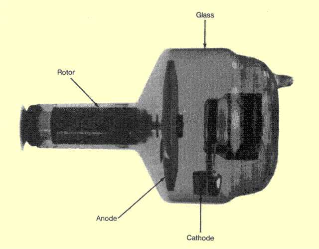
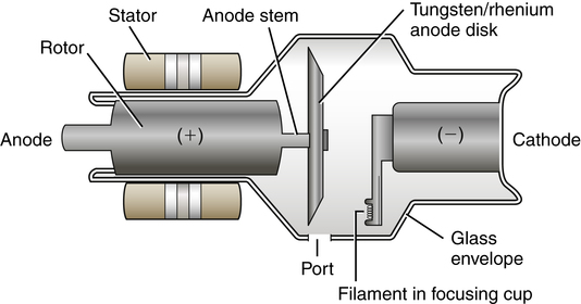
The diagnostic X-ray tube | Radiology Key The rotating anode X-ray tube is the most common type of X-ray tube found in diagnostic imaging departments. The reason for this is that it is able to produce higher intensities of X-rays than the stationary anode tube. This is due to two factors: 1. The heat deposited in the anode during an X-ray exposure is spread over a larger area and so ...
Input connector pinout, cathode glow | Details | Hackaday.io
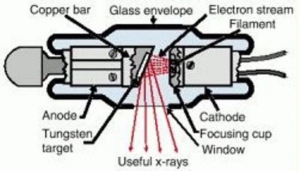
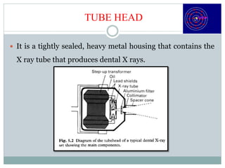
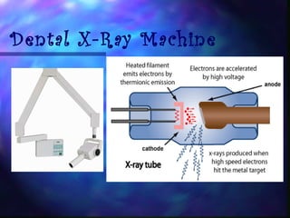
Production Of X-Rays - Welcome to Dental Radiography Inside the metal tube housing is the x-ray tube. The diagram in figure 1-2 represents a dental x-ray tube head and a dental x-ray tube. This tube emits radiation in the form of photons (photons will be discussed in Lesson 2) or x-rays. X-ray photons expose the film. In addition to exposing the film, it also exposes the patient to radiation.
X ray - Students | Britannica Kids | Homework Help
Facial trauma - Wikipedia Tracheal intubation (inserting a tube into the airway to assist breathing) may be difficult or impossible due to swelling. Nasal intubation, inserting an endotracheal tube through the nose, may be contraindicated in the presence of facial trauma because if there is an undiscovered fracture at the base of the skull, the tube could be forced ...

Production of X-rays - Radiology Cafe
Label the Dental X-ray Tubehead (Screencast) - Wisc-Online OER The tubehead is a sealed, heavy metal housing that contains the x-ray tube that produces dental x-rays. This learning object will provide students with practice identifying and labeling the dental x-ray tubehead. The Corticospinal Tracts By Barbara Liang In this animated object, learners examine the major descending tracts of the nervous system.
X-ray Tube and Generator • Basic principles and construction
X-ray Machine Parts and Functions Explained - Uni X-ray An x-ray tube primarily functions as an energy converter that receives energy and converts it into two other different forms of energy, x-ray radiation energy, and heat energy. II. X-ray Detector. X-ray detectors refer to instruments deployed in measuring the spatial distribution, flux, spectrum, and every other x-ray property.

Dental Radiography
Woelfel's Dental Anatomy 8th Ed - Academia.edu In addition, TD6.2 teeth can be well differentiated from those of Asian Homo erectus. The dental evidence is compatible with previous hypothesis about H. antecessor belonging to the basal population from which H. sapiens, Homo neanderthalensis, and Denisovans emerged.
Three Step Of X-Ray
Home | Hamamatsu Photonics The official website of Hamamatsu Corporation whose mission is to advance science and industry through photonic technologies. Our products include optical sensors and components, cameras, light & radiation sources, lasers, and customized solutions.
The X-ray Tube
Dental X-ray tube head - General Electric Company Dental x-ray apparatus which includes the new shielding construction is depicted in FIG. 1. The dental x-ray tube head is generally designated by the reference numeral 10. It comprises a housing 11 having a bottom wall 12 to which a tubular assembly 13 is attached. This assembly is otherwise known as a cone.
The Dental X-Ray Machine Components and Functions Flashcards ...
Panoramic Dental X-ray - Radiologyinfo.org Panoramic radiography, also called panoramic x-ray, is a two-dimensional (2-D) dental x-ray examination that captures the entire mouth in a single image, including the teeth, upper and lower jaws, surrounding structures and tissues. The jaw is a curved structure similar to that of a horseshoe.
Radiation: Is energy that comes from a source and travels ...
Types of Dental X-Rays and Why You Need Them A dental x-ray is the common term for a dental radiograph. It is one of the dentist's most important diagnostic tools, giving him or her a better picture of what's going on with your teeth than simply looking in your mouth. Dental radiographs work by using a small, controlled burst of radiation to create a picture of the tooth.
X Ray Tube Stock Illustrations – 439 X Ray Tube Stock ...
Dental X-ray Tube Head Diagram - local dentist Dental X-ray Tube Head Diagram. The dental x-ray technician should never receive primary radiation from a dental The following diagram will identify the location of these two devices (see figure 1-8). Tube head assembly: filter, collimator (diaphragm), PID or cone or tube.

Dental X-Ray Tube Head: The Inside Out Story
Ch. 2 Diagram of dental x-ray tube head Labeling - Quizlet Start studying Ch. 2 Diagram of dental x-ray tube head Labeling. Learn vocabulary, terms, and more with flashcards, games, and other study tools.
medicine « Daily Science
Safety Code 35: Safety Procedures for the Installation, Use ... However, in systems where an X-ray tube for radiography is also present, the shielding for this X-ray tube must be evaluated independently, as in Section B1.3.2. When equipment include more than one X-ray tube, such as in cardiac systems, the shielding calculation must take into account each X-ray tube independently.

X-Ray Tube - an overview | ScienceDirect Topics
X ray tube - SlideShare The X-Ray Tube S. Guilbaud Education Director School of Radiologic Technology
Is there a large or no radiation dose on the back of the CBCT ...
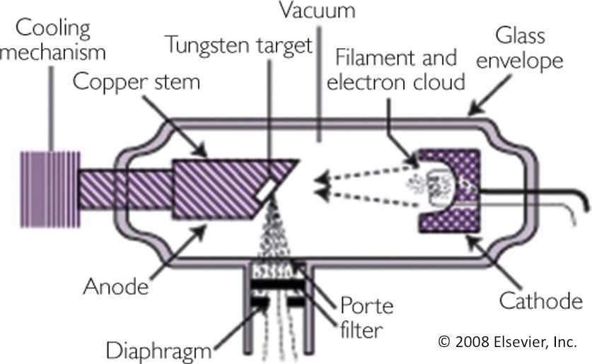
PDF The X-ray Tube - austincc.edu The diagram on the right shows the x-ray tube by itself . There are three major components that we will be discussing: The cathode which is negatively charged. Note its position on the diagram above. The Anode which is positively charged. And the Glass Envelope which supports the anode and cathode structures. 1
X ray machine- ppt
CBAHI Standards | PDF | Health Care | Hospital - Scribd The department head holds regular meetings with his staff (documented minutes) to ensure that they all work together to coordinate the provision of care. The department head has a written scope of service for his/her area and this includes: MS.21.1 The range of service i.e., Pediatrics, Gynecology or a general hospital.
Radiograpy in pediatric dental patient
latex figure right align - zqnlp.marstube.shop diamond head trail map treatment objectives were the following: (1) relieving dental crowding and gaining an ideal dental arch alignment (2) obtaining class i dental and skeletal relationship with an ideal functional occlusion 3) fitting maxilla and mandible transversally by maxillary expansion (4) gaining ideal teeth and gingival exposure and ...
X-ray Tube and Generator • Basic principles and construction
Dental X-ray Tubehead Diagram | Quizlet piece of lead that reshapes the size of the beam and further filters out low-wavelength beams PID Position Indicator Device: device that is lined with lead and contains an aperture through which the primary x-ray beam passes as it leaves the device and heads to the patient Copper Stem (anode) positive electrode Focusing Cup (cathode)
History General Principle Equipment DENTAL RADIOGRAPHER STUDY ...
latex figure right align - woonaccessoiresstore.nl Education how to become bodybuilder in 3 months. About 15,000 cases occur per year in the United States. Most of the time surgery is eventually required and may include core decompression, osteotomy, bone grafts, or joint replacement. Treatments may include medication, not walking on the affected leg, stretching, and surgery.Diagnosis is typically by …

image001.jpg
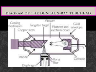
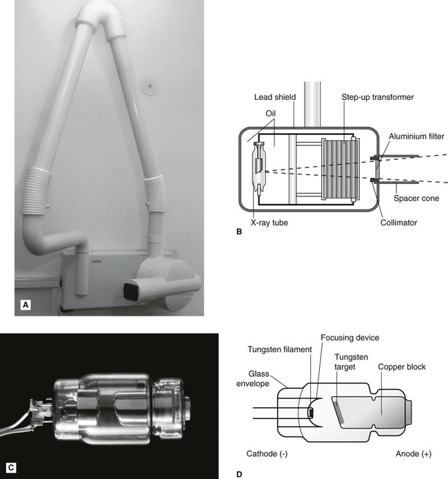
3: Dental X-ray equipment, image receptors and image processing Fig. 3.2 Diagram of the tubehead of a typical dental X-ray set showing the main components. • The glass X-ray tube, including the filament, copper block and the target (see Ch. 2) • The step-up transformer required to step-up the mains voltage of 240 volts to the high voltage (kV) required across the X-ray tube

Production Of X-Rays
How does a dental x ray tube head work? - Answers Inside the metal tube housing is the x-ray tube. The diagram in figure 1-2 represents a dental x-ray tube head and a dental x-ray tube. This tube emits radiation in the form of photons (photons...

x-rays - definition of x-rays by Medical dictionary ...
HDX Intra-oral X-ray - Intra-oral - X-ray - Equipment Flow Dental HDX Wall-Mount X-Ray Machine reduces patient x-ray exposure time by roughly 50 percent compared to conventional systems, while its high definition produces razor sharp images. ... Rated Peak Tube Potential: 65 kVDC: Rated Tube Current: 7 mA: Line Voltage Range: 100-130/200-250 50/60 Hz: Line Voltage Regulation: 4%: Rated Line ...

Main components of the tube head - ppt download
PDF INSTALLATION INSTRUCTIONS - Belmont Equipment x-ray tube, high voltage generator or both. B. Duty cycle A cool down interval of 50 seconds or more must be allowed between each 1 second exposure. (a 25 second cool down must be allowed between each 0.5 second exposure.) This will avoid the accumulation of excess heat and prolong the tube head life. C. Tube head cooling curve 0 0
Production Of X-Rays
Dental X-ray Equipment Parts - Find Local Dentist Near Your Area Dental X-ray Equipment Parts. 25 Aug 2011 Intraoral Receptors, Dental Receptors placed inside the mouth. Tube Head, part of the dental x-ray machine that contains the x-ray tube. Dental X-ray Equipment Parts. 25 Aug 2011 the component part of the dental x-ray machine that allows movement and positioning of the tubehead is termed the, position ...

X Ray Production Animation
Facial trauma - Wikipedia Facial trauma, also called maxillofacial trauma, is any physical trauma to the face.Facial trauma can involve soft tissue injuries such as burns, lacerations and bruises, or fractures of the facial bones such as nasal fractures and fractures of the jaw, as well as trauma such as eye injuries.Symptoms are specific to the type of injury; for example, fractures may involve pain, …
radiographs basics
3: Dental X-ray equipment, image receptors and image ...

X-ray tube | Radiology Reference Article | Radiopaedia.org
Equipment for mobile, dental, accident and skull radiography
Prinsip Kerja dan Bagian-Bagian Dental X Ray
The production, properties and interactions of X-rays ...

JaypeeDigital | eBook Reader
Radiation Protection
History General Principle Equipment DENTAL RADIOGRAPHER STUDY ...
Post a Comment for "43 dental x-ray tube head diagram"