43 draw the human heart and label its parts
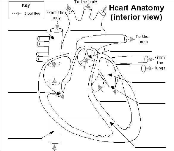
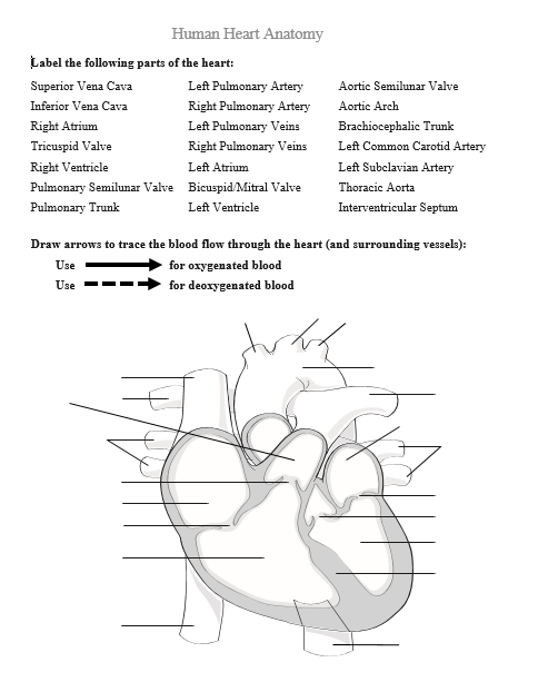
Heart Anatomy: Labeled Diagram, Structures, Blood Flow ... - EZmed There are 4 chambers, labeled 1-4 on the diagram below. To help simplify things, we can convert the heart into a square. We will then divide that square into 4 different boxes which will represent the 4 chambers of the heart. The boxes are numbered to correlate with the labeled chambers on the cartoon diagram. View fullsize draw the diagram of sectional view of human heart and on it name and ... answered draw the diagram of sectional view of human heart and on it name and label the following parts : (a) the Chamber of the heart that pumps out deoxygenated blood (b) the blood vessel that carries away oxygenated blood from the heart . (c) the blood vessel that receives deoxygenated blood from the lower part of our body . Answer 4.6 /5 103
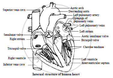
Draw a diagram to show the internal structure of the human heart. Label ... Label six parts in all including two valves at least. Advertisement Expert-verified answer ikrakhadim Here is your answer! The human heart consists of many parts, so this diagram will help you out. It involves; Artery Aorta Vena cava Ventricle and any more. Advertisement Answer Moongirl1 Answer: Ok See in the above picture

Draw the human heart and label its parts
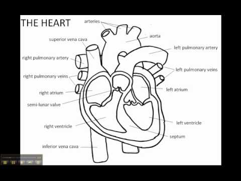
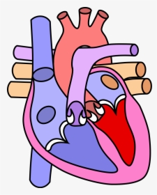
How to Draw the Internal Structure of the Heart (with Pictures) - wikiHow To draw the internal structure of a human heart, follow the steps below. Part 1 Finding a Diagram 1 To find a good diagram, go to Google Images, and type in "The Internal Structure of the Human Heart". Find an image that displays the entire heart, and click on it to enlarge it. 2 Find a piece of paper and something to draw with. Easy trick to draw Human Heart - YouTube This is how you can draw human heart in very easy way, stay tuned for more videos like this. A Labeled Diagram of the Human Heart You Really Need to See The human heart, comprises four chambers: right atrium, left atrium, right ventricle and left ventricle. The two upper chambers are called the left and the right atria, and the two lower chambers are known as the left and the right ventricles. The two atria and ventricles are separated from each other by a muscle wall called 'septum'.
Draw the human heart and label its parts. Draw a diagram of the human heart and label its parts. - Science Draw a diagram of the human heart and label its parts. CBSE CBSE (English Medium) Class 10. Question Papers 892. Textbook Solutions 20383. MCQ Online Tests 12. ... Draw a diagram of the human heart and label its parts. Advertisement Remove all ads. Solution Show Solution. Diagram of human heart: Concept: Human Heart. Parts Of The Human Heart | Science Trends The parts of the human heart can be broken down into four chambers, muscular walls, vessels, and a conductive system. The two upper chambers are called the atria, with lower parts called ventricles. These all work together to make up the vital function of your heart. Everybody knows that the human heart is the essential organ in our bodies. Human Heart Diagram - Human Body Pictures - Science for Kids Find free pictures, photos, diagrams, images and information related to the human body right here at Science Kids. Photo name: Human Heart Diagram Picture category: Human Body Image size: 70 KB Dimensions: 600 x 600 Photo description: This is an excellent human heart diagram which uses different colors to show different parts and also labels a number of important heart component such as the ... Draw the structure of human heart and label its parts - Biology Draw the structure of human heart and label its parts . CISCE ICSE Class 6. Textbook Solutions 7180. Question Bank Solutions 6879. Concept Notes & Videos 203 ... Draw the structure of human heart and label its parts. Advertisement Remove all ads. Solution Show Solution. Concept: Blood Circulatory System in Human ...
Label the heart — Science Learning Hub In this interactive, you can label parts of the human heart. Drag and drop the text labels onto the boxes next to the diagram. Selecting or hovering over a box will highlight each area in the diagram. pulmonary vein semilunar valve right ventricle right atrium vena cava left atrium pulmonary artery aorta left ventricle Download Exercise Tweet How to Draw a Human Heart: 11 Steps (with Pictures) - wikiHow Sketching the Heart 1 Draw the lower half of an acorn shape so it's tilted to the left. Use your pen or pencil to start drawing the main part of the heart. This should look like an open-ended acorn that's missing its cap. Draw the shape so it's tilted about 120 degrees to the left. [1] Heart Diagram - 15+ Free Printable Word, Excel, EPS, PSD Template ... This is a heart diagram template representing a heart and its parts. It is a vintage picture taken from an old medical book which can easily be used for studying the details of a heart. Anatomical Heart Diagram of Blue Baby This is a heart diagram template clearly showing the blue baby syndrome, which is caused due to decrease of oxygen in babies. Draw the diagram of the sectional view of the human heart and label the ... Biology. Grade 10. Heart. Answer. Draw the diagram of the sectional view of the human heart and label the following parts. A) The chambers of the heart that pumps out deoxygenated blood. B) The blood vessel that receives deoxygenated blood from the other parts of the body. C) The blood vessel that carries away oxygenated blood from the heart.
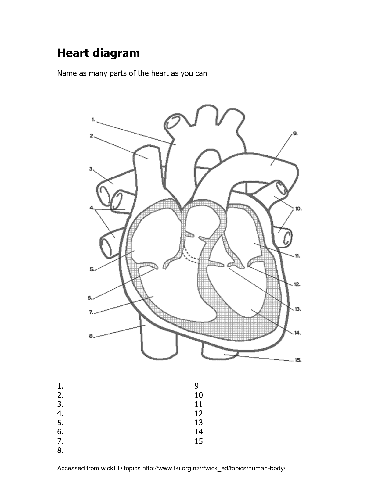
Human Heart - Anatomy, Functions and Facts about Heart - BYJUS Following are the main functions of the heart: One of the primary functions of the human heart is to pump blood throughout the body. Blood delivers oxygen, hormones, glucose and other components to various parts of the body, including the human heart. The heart also ensures that adequate blood pressure is maintained in the body. Human Heart - Diagram and Anatomy of the Heart - Innerbody The heart contains 4 chambers: the right atrium, left atrium, right ventricle, and left ventricle. The atria are smaller than the ventricles and have thinner, less muscular walls than the ventricles. The atria act as receiving chambers for blood, so they are connected to the veins that carry blood to the heart. Heart Diagram with Labels and Detailed Explanation - BYJUS Diagram of Heart. The human heart is the most crucial organ of the human body. It pumps blood from the heart to different parts of the body and back to the heart. The most common heart attack symptoms or warning signs are chest pain, breathlessness, nausea, sweating etc. The diagram of heart is beneficial for Class 10 and 12 and is frequently ... The Human Heart || How to Draw Human Heart in Very Easy Step || by ... This is my second video on the Human heart based on general and previous knowledge which we have been reading for years. please go step by step as I am teach...
How to draw human heart
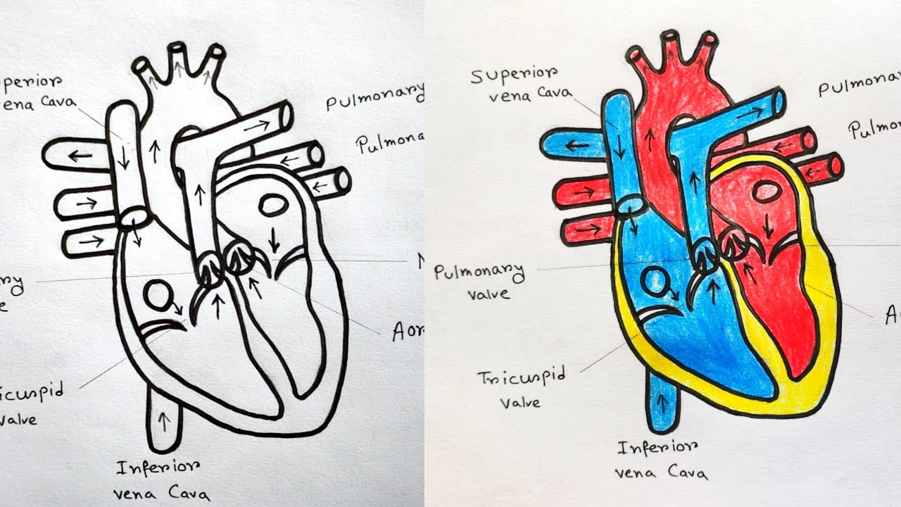
Draw the diagram of sectional view of human heart and label the ... - Toppr Draw the diagram of sectional view of human heart and label the following parts a. The chambers of the heart that pumps out deoxygenated blood. b. The blood vessel that receives deoxygenated blood from the other parts of the body. c. The blood vessel that carries away oxygenated blood from the heart. d. Part which prevents the back flow of blood.

Draw a diagram of the human heart and label its parts.
The 18 parts of the human heart, and their functions 9. Left ventricle. The left ventricle contains the strongest muscles in the whole heart. From this ventricle, blood is pumped into the aortic artery, which divides to water the rest of the body's blood. The blood pressure generated by this ventricle must be much higher than that generated by the right ventricle. 10.

How to Draw the Human Heart and Label Its Parts – Draw Swan
13 parts of the human heart (and its functions) - LORECENTRAL In general we can find the following parts of the heart . 1. Left atrium. One of the four main heart cavities in which blood is received and pumped . The left atrium is characterized by being connected to the pulmonary veins, from which it receives highly oxygenated blood and then sends it to the left ventricle. 2.

How to Draw a Human Heart: 11 Steps (with Pictures) - wikiHow
Human Heart (Anatomy): Diagram, Function, Chambers, Location in Body The heart is a muscular organ about the size of a fist, located just behind and slightly left of the breastbone. The heart pumps blood through the network of arteries and veins called the ...
With the Help of a Labelled Diagram Describe the Internal ...
Prepare a drawing of the human heart and label its parts. | Quizlet Find step-by-step Physical science solutions and your answer to the following textbook question: Prepare a drawing of the human heart and label its parts.. ... Identify the parts of the human heart, and describe the function of each part. PHYSICS. Estimate the number of times a human heart beats during its lifetime.

Amazon.com: Lunarable Human Heart Throw Blanket, Anatomic ...
Human Heart Diagram Labeled | Science Trends The left ventricle and left atrium make up the left heart while the right ventricle and right atrium make up the right heart. While there are four different chambers of the heart, the chambers work together and the heart basically functions as a single organ. The muscle tissue dividing the two halves of the heart is referred to as the septum.

Q1 Given alongside is a diagram of human heart showing its ...
Draw a diagram of the front view of human heart and label any six parts ... Draw a diagram of the front view of human heart and label any six parts including at least two, that are concerned with arterial blood supply to the heart muscles. life processes; ... Draw a sectional view of the human heart and label on it - Aorta, Right ventricle and Pulmonary veins. asked Oct 12, 2019 in Biology by Suchita (66.6k points ...

How to Draw Human Heart – 7 easy steps
File:Diagram of the human heart (cropped).svg - Wikipedia Added shadows. Left main pulmonary artery with its first division. 07:02, 2 June 2006: 650 × 650 (26 KB) Yaddah: Diagram of the human heart, created by Wapcaplet in Sodipodi. Cropped by ~~~ to remove white space (this cropping is not the same as Wapcaplet's original crop). == See also == * Image:Diagram of the human heart.svg - original
A schematic diagram of a longitudinal section of the human ...
Diagram of Human Heart and Blood Circulation in It A heart diagram labeled will provide plenty of information about the structure of your heart, including the wall of your heart. The wall of the heart has three different layers, such as the Myocardium, the Epicardium, and the Endocardium. Here's more about these three layers. Epicardium

Q4 Given alongside is a diagram of the human heart showing ...
A Diagram of the Heart and Its Functioning Explained in Detail Human heart is covered by a double layered structure which is known as pericardium. The outer layer is associated with the major blood vessels whereas the inner layer is attached to the cardiac muscles. These layers are separated by a pericardial fluid. This covering is like a membrane which holds all the parts of the heart. Chambers
Draw the diagram showing the sectional view of the human ...
Draw a labelled diagram of Human Heart. Draw a table to show the ... Click here👆to get an answer to your question ️ Draw a labelled diagram of Human Heart. Draw a table to show the functions of any two chambers of Human Heart. Solve Study Textbooks ... The Fish Tale Across the Wall Tenths and Hundredths Parts and Whole Can you see the Pattern? class 6. Maps Practical Geometry Separation of Substances ...
Free Human Heart Sketch Diagram, Download Free Human Heart ...
A Labeled Diagram of the Human Heart You Really Need to See The human heart, comprises four chambers: right atrium, left atrium, right ventricle and left ventricle. The two upper chambers are called the left and the right atria, and the two lower chambers are known as the left and the right ventricles. The two atria and ventricles are separated from each other by a muscle wall called 'septum'.

Colorful Hand Drawn Illustration Of Human Heart Anatomy Stock ...
Easy trick to draw Human Heart - YouTube This is how you can draw human heart in very easy way, stay tuned for more videos like this.
Free Human Heart Sketch Diagram, Download Free Human Heart ...
How to Draw the Internal Structure of the Heart (with Pictures) - wikiHow To draw the internal structure of a human heart, follow the steps below. Part 1 Finding a Diagram 1 To find a good diagram, go to Google Images, and type in "The Internal Structure of the Human Heart". Find an image that displays the entire heart, and click on it to enlarge it. 2 Find a piece of paper and something to draw with.

How to Draw The Human Heart Diagram with Label Easily? Get Step by Step Guidance of Artist.

Heart Structure | BioNinja

How to Draw a Human Heart: 11 Steps (with Pictures) - wikiHow
heart | Structure, Function, Diagram, Anatomy, & Facts ...
Draw the diagram showing the sectional view of the human ...
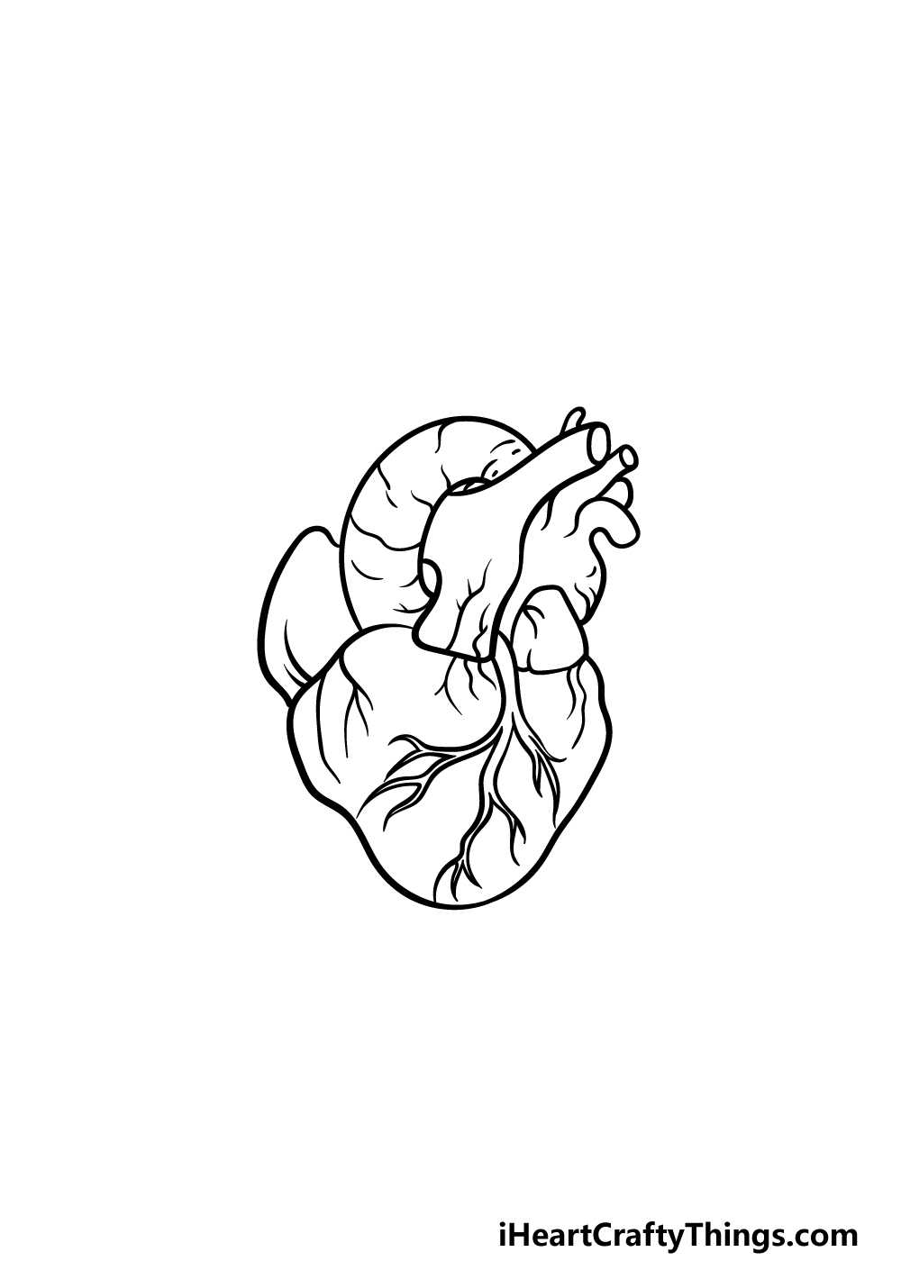
Human Heart Drawing - How To Draw A Human Heart Step By Step

Draw the diagram of sectional view of human heart and on it ...
13+ Heart Diagram Templates – Sample, Example, Format ...

How to draw human heart labeled | Human drawing, Biology ...

Draw a diagram of the human heart and label its parts ...

Right Ventricle - Heart - Human Body - Education Stock ...

Human heart drawing. An image of a human heart drawing ...
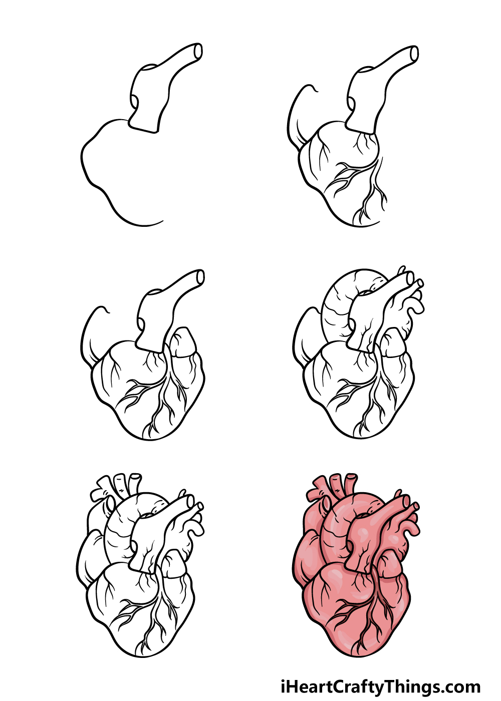
Human Heart Drawing - How To Draw A Human Heart Step By Step
How to draw Human Heart with colour | Human Heart labelled diagram|Human Heart drawing easy tutorial
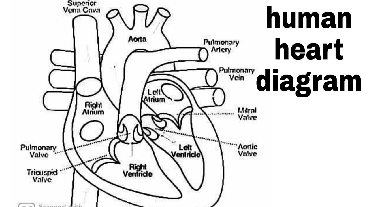
Diagram of heart/How to draw human heart easily/human heart/human heart diagram/labelled human heart

Human Heart for Kids: 2 Fun Heart Models plus Worksheets
Draw a diagram of the front view of human heart and label any ...
13+ Heart Diagram Templates – Sample, Example, Format ...
Human heart hi-res stock photography and images - Alamy

How to Draw a Human Heart: 11 Steps (with Pictures) - wikiHow
Solved Aortic Semilunar Valve Human Heart Anatomy Label the ...
Draw the structure of a human heart and label its parts ...

Heart Anatomy: Labeled Diagram, Structures, Blood Flow ...
heart structure with name - Clip Art Library

File:Diagram of the human heart (cropped).svg - Wikimedia Commons

4,068 Human Heart Diagram Stock Photos, Pictures & Royalty ...

How to Draw a Human Heart: 11 Steps (with Pictures) - wikiHow
Clip Art Library Download Clipart Human Heart - Longitudinal ...

draw the diagram of human heart and label the parts which:(i ...
Post a Comment for "43 draw the human heart and label its parts"