38 microscope labeled worksheet
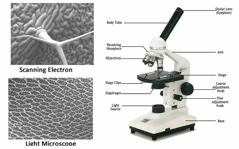
CELLS alive! Since 1994, CELLS alive! has provided students with a learning resource for cell biology, microbiology, immunology, and microscopy through the use of mobile-friendly interactive animations, video, puzzles, quizzes and study aids. Tissues Class 9 ppt - [PPTX Powerpoint] - VDOCUMENT Aug 17, 2015 · 4. Remove this peel and put it in a Petri dish filled with water and add a few drops of safranin. 5. Wait for few minutes and then transfer it onto a slide. Gently place a cover slip over it and observe under microscope. When observed under microscope,outermost layer of cells called EPIDERMIS (epidermal tissue) is seen.
UD Virtual Compound Microscope - University of Delaware ©University of Delaware. This work is licensed under a Creative Commons Attribution-NonCommercial-NoDerivs 2.5 License.Creative Commons Attribution-NonCommercial-NoDerivs 2.5 License.
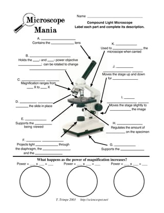
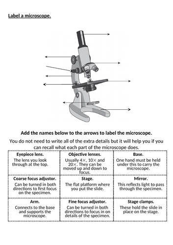
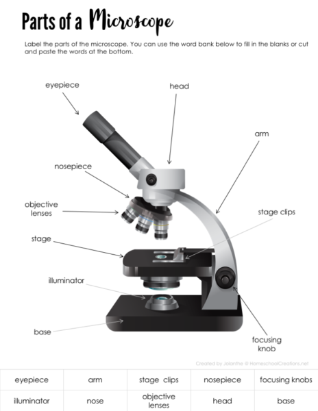
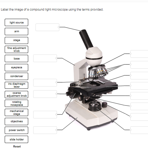
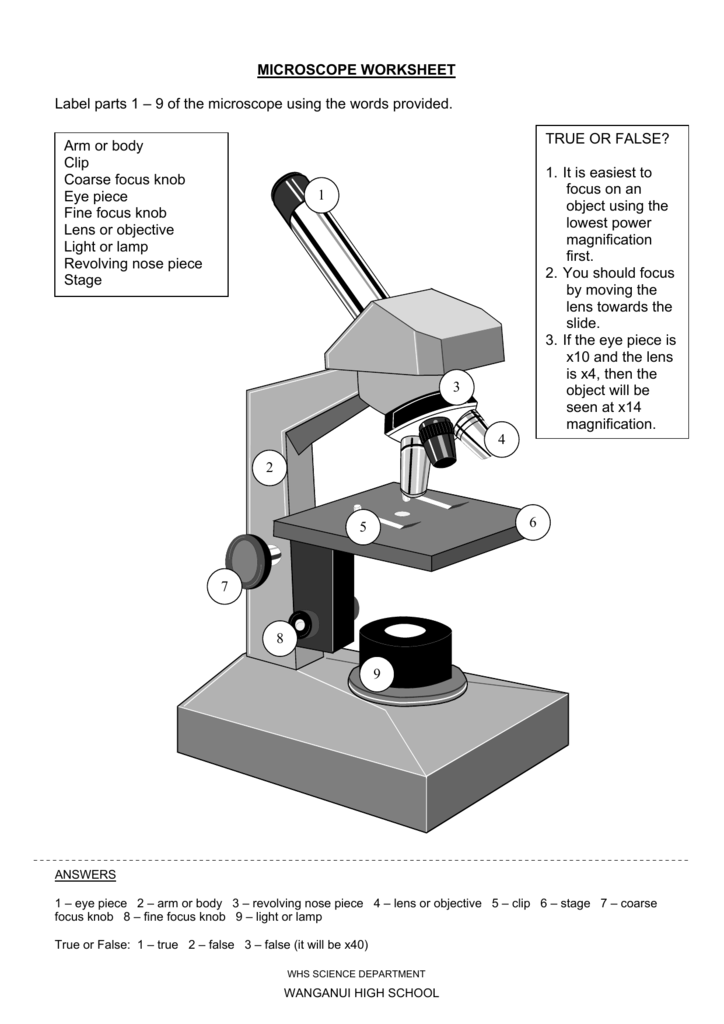
Microscope labeled worksheet
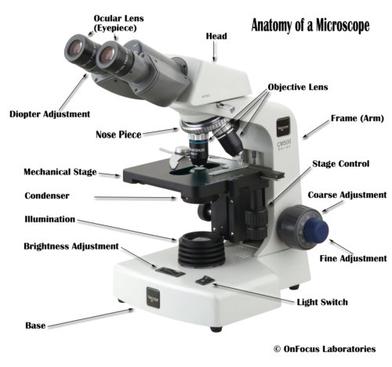
Parts of a microscope with functions and labeled diagram Sep 17, 2022 · Q. Differentiate between a condenser and an Abbe condenser. Ans. Condensers are lenses that are used to collect and focus light from the illuminator into the specimen. They are found under the stage next to the diaphragm of the microscope. They play a major role in ensuring clear sharp images are produced with a high magnification of 400X and above. Interactive Bacteria Cell Model - CELLS alive Periplasmic Space: This cellular compartment is found only in those bacteria that have both an outer membrane and plasma membrane (e.g. Gram negative bacteria).In the space are enzymes and other proteins that help digest and move nutrients into the cell. Cell Wall: Composed of peptidoglycan (polysaccharides + protein), the cell wall maintains the overall shape of a … Science — Biology – Easy Peasy All-in-One Homeschool Make observations. (If you have a microscope, you can rub some skin off of you and look at it with your microscope.) ... Add this worksheet to your portfolio. ... Make sure it is labeled clearly so that someone can understand it without you explaining it to them. Lesson 178. Write your conclusion. Older students should write a paragraph. Lesson ...
Microscope labeled worksheet. Plant Cell- Definition, Structure, Parts, Functions, Labeled Diagram Sep 16, 2022 · Figure: Labeled diagram of plant cell, created with biorender.com. The typical characteristics that define the plant cell include cellulose, hemicellulose and pectin, plastids which play a major role in photosynthesis and storage of starch, large vacuoles responsible for regulating the cell turgor pressure. Join LiveJournal Password requirements: 6 to 30 characters long; ASCII characters only (characters found on a standard US keyboard); must contain at least 4 different symbols; Gel Electrophoresis - University of Utah Have you ever wondered how scientists work with tiny molecules that they can't see? Here's your chance to try it yourself! Sort and measure DNA strands by running your own gel electrophoresis experiment. Parts of Stereo Microscope (Dissecting microscope) – labeled … Unlike a compound microscope that offers a flat image, stereo microscopes give the viewer a 3-dimensional image that you can see the texture of a larger specimen. [In this image] Examples of Stereo & Dissecting microscopes. Major microscope brands (Zeiss, Olympus, Nikon, Amscope, Omano, Leica …) all produce stereomicroscopes.
Science — Biology – Easy Peasy All-in-One Homeschool Make observations. (If you have a microscope, you can rub some skin off of you and look at it with your microscope.) ... Add this worksheet to your portfolio. ... Make sure it is labeled clearly so that someone can understand it without you explaining it to them. Lesson 178. Write your conclusion. Older students should write a paragraph. Lesson ... Interactive Bacteria Cell Model - CELLS alive Periplasmic Space: This cellular compartment is found only in those bacteria that have both an outer membrane and plasma membrane (e.g. Gram negative bacteria).In the space are enzymes and other proteins that help digest and move nutrients into the cell. Cell Wall: Composed of peptidoglycan (polysaccharides + protein), the cell wall maintains the overall shape of a … Parts of a microscope with functions and labeled diagram Sep 17, 2022 · Q. Differentiate between a condenser and an Abbe condenser. Ans. Condensers are lenses that are used to collect and focus light from the illuminator into the specimen. They are found under the stage next to the diaphragm of the microscope. They play a major role in ensuring clear sharp images are produced with a high magnification of 400X and above.
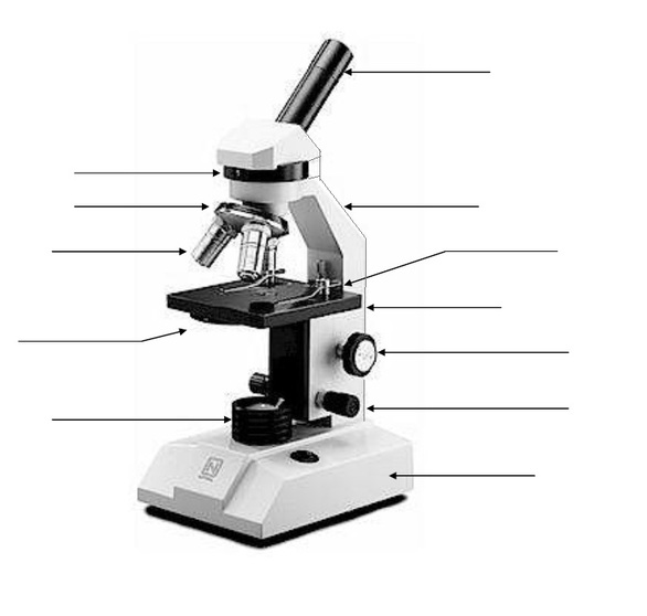
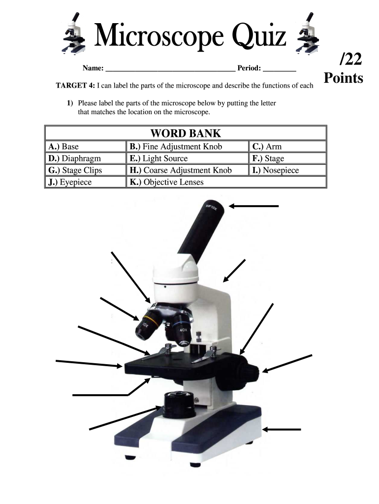
Microscope parts
Labeled Parts Of A Microscope - ClipArt Best

Copy of Compound Microscope Worksheet.pdf - Biology I LCHS ...

Compound Microscope- Definition, Labeled Diagram, Principle ...

BIOS135_W4_Microscope_Lab_Worksheet.docx - Microscope Lab ...
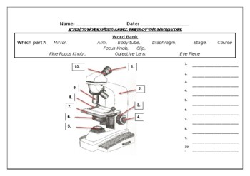
Science worksheet: Label The Parts Of A Microscope by Science ...
Microscope Parts Quiz

Label Compound Microscope Lesson Plans & Worksheets

Label a Microscope Worksheet

Microscope Diagram Labeled, Unlabeled and Blank | Parts of a ...

SOLVED: Directions: Label the microscope below: Nto: Identify ...

Free Labeling Scientific Tools (Microscope)/4

Microscope Parts Practice Worksheet

Label the microscope — Science Learning Hub
7Ac Microscope Labelling Worksheet | Teaching Resources

Biology label part of microscope

Parts of a Microscope Labeling Activity

Parts of a Microscope worksheet
Lab Station: Parts of the Microscope

School | Reading worksheets, Microscope parts, Biology lessons

13 - Microscope Parts - PowerPoint Worksheet.docx - 1 Name: _ ...

Parts of a Microscope with Their Functions – Microbe Online
Parts of a Microscope - Free Printable

Microscope worksheet

Parts of the Microscope worksheet
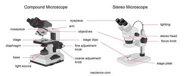
Parts of Stereo Microscope (Dissecting microscope) – labeled ...
Solved PLEASE HELP THANK YOU- LIGHT SOURCE WILL BE NUMBER 1 ...
Free Microscope Worksheets for Simple Science Fun for Your ...

KS3&4 Parts of a Light Microscope Worksheet | Teaching Resources

Microscope Diagram - Free Printable Tests and Worksheets ...

Microscope Maintenance Tips | Science supplies, Microscope ...
Microscope Parts and Functions
microscope | The Biology Corner
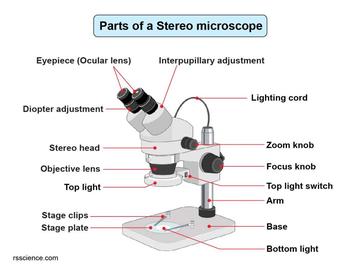
Parts of Stereo Microscope (Dissecting microscope) – labeled ...
Microscope Worksheet
Microscope Fill In The Blank - Fill Online, Printable ...
LAB 1: Scientific Method/Tools of Scientific Inquiry
Free Microscope Worksheets for Simple Science Fun for Your ...
Post a Comment for "38 microscope labeled worksheet"