39 cow eye diagram labeled
eye diagram labeled - anatomychart.z21.web.core.windows.net brain function structure functions diagram lobes macmillan labelled showing. Cow Eye Dissection - YouTube . cow eye dissection. Label The Muscles Of The Eye - PurposeGames . purposegames. 3d Eye Model 32 Pcs Assembled Human Anatomy Model New 3D Structure Of . auge. Photoreceptor Cell ... Cow's Eye Dissection | Exploratorium Learn how to dissect a cow's eye in your classroom. This resource includes: a step-by-step, hints and tips, a cow eye primer, and a glossary of terms.
Cow Eye Labeled Diagram - ClipArt Best 31 cow eye labeled diagram. Free cliparts that you can download to you computer and use in your designs.
Cow eye diagram labeled
eye diagram labeled - anatomyhealth.z21.web.core.windows.net brain function structure functions diagram lobes macmillan labelled showing. Cow Eye Dissection - YouTube . cow eye dissection. Label The Muscles Of The Eye - PurposeGames . purposegames. 3d Eye Model 32 Pcs Assembled Human Anatomy Model New 3D Structure Of . auge. Photoreceptor Cell ... Cow Eye Dissection & Parts of the Eye Diagram | Quizlet Located in the back of the eye, contains the rods and cones. pupil The opening in the center of the iris through which light enters the eye lens The transparent structure behind the pupil that changes shape to help focus images on the retina. ligaments (eye) Fibers that connect ciliary muscles to the lens in the eye blind spot (optic disc) Cow Anatomy - External Body Parts and Internal Organs with Labeled Diagram I will try to show you all of the body parts of a cow in labeled diagrams. The external body parts from the head region of a cow - in this head region, you might identify the mouth, lip, cheek, chin, muzzle, forehead, poll, ear, eye, nostril, and other.
Cow eye diagram labeled. Herpes simplex virus 1 (HSV-1)- An Overview - Microbe Notes Aug 25, 2022 · Source: Wikipedia. Invasion of cells by HSV1 requires binding of the envelope gC (glyco-protein-C) and/or gB to Heparan sulfate receptors, engagement by gD of one of several co-receptors including HveA (Herpes virus entry mediator A, also known as HVEM, Herpes Virus Entry Mediators), fusion of the viral envelope with the cell plasma membrane and delivery of … Cow's Eye Dissection - step 2 - Exploratorium Look all around. Six muscles attached to your eyeball move your eye so you can look in different directions. Cows have only four muscles that control their eyes ... Cow Eye Dissection | Carolina.com Jul 24, 2013 — Explore the internal and external anatomy of the cow eye with this dissection. It can be carried out in one or two class periods and ... labelled diagram of a cow A Diagram Of The Body Parts Of A Cow | 4-H Project: Livestock . cow parts body diagram cattle cows animal veterinarian. Cow . cow labeling diagrams pdf resolution exploringnature. Cow's Eye Diagram Quiz . eye diagram cow quiz cows purposegames. Labeled Volvox Diagram - Made By Creative ...
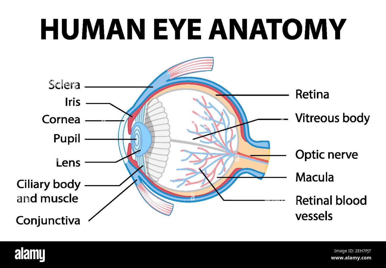
Join LiveJournal Password requirements: 6 to 30 characters long; ASCII characters only (characters found on a standard US keyboard); must contain at least 4 different symbols; Duck Anatomy - External and Internal Features with Labeled Diagram ... Jul 24, 2021 · Eye, ear, and nape of a duck; Neck, flank, and abdomen of a duck; Mantle, back, and wing of a duck; Tail and uropygial or preen gland of duck; Shank, feet, and toe of a duck; The keel of a duck; Different types of feathers (wing flight feathers, flight tail feathers, coverts, and more) You will find all the features of the duck’s body in the ... Cow Eye Dissection & Anatomy Project | HST Learning Center Cow Eye Dissection: Internal Anatomy 1. Place the cow's eye on a dissecting tray. The eye most likely has a thick covering of fat and muscle tissue. Carefully cut away the fat and the muscle. As you get closer to the actual eyeball, you may notice muscles that are attached directly to the sclera and along the optic nerve. human eye diagram labeled visual system anatomy understanding. Optics: Eye Physiology alchemical.org. eye eyes physiology anatomy diagram optics. Cow's Eye Dissection - Eye Diagram . eye cow diagram blank dissection parts exploratorium label eyes worksheet science human clipart edu learning cliparts clip body library primer
Cow Eye Labeled - All About Cow Photos - votenickpang.com Cow S Eye Dissection Diagram Solved 6 The Images Below Show A Preserved Cow S Eye One Chegg Neur 320 Art And Vision Ppt Cow Eye Dissection Powerpoint Ation Id 3482425 Labeling Cow Eye Diagram Quizlet Diagram Of Human Eye Anatomy With Label Stock Ilration 74745446 Pixta Course Help Online - Have your academic paper written by a … We will take care of all your assignment needs. We are a leading online assignment help service provider. We provide assignment help in over 80 subjects. eye diagram labeled - anatomyedu99.z21.web.core.windows.net brain function structure functions diagram lobes macmillan labelled showing. Cow Eye Dissection - YouTube . cow eye dissection. Label The Muscles Of The Eye - PurposeGames . purposegames. 3d Eye Model 32 Pcs Assembled Human Anatomy Model New 3D Structure Of . auge. Photoreceptor Cell ... Anatomy- Cow Eyeball Diagram | Quizlet Start studying Anatomy- Cow Eyeball. Learn vocabulary, terms, and more with flashcards, games, and other study tools. ... cow eye. 15 terms. madison_cooke5. ECG. 12 terms. cynnat PLUS. Other sets by this creator. Anatomy- Histology. 11 terms. alyssatuttobene. Anatomy- Body Vocab 4. 11 terms.
Retinal Detachment | Health and Nutrition Facts for You ...
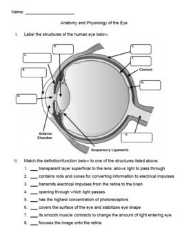
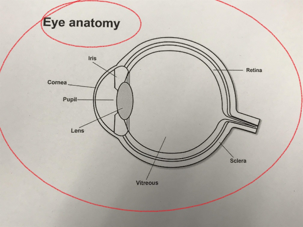
Cow Eye Dissection Flashcards | Quizlet A ring of muscle tissue that forms the colored portion of the eye around the pupil and controls the size of the pupil opening. Lens The transparent structure behind the pupil that changes shape to help focus images on the retina. Retina Located in the back of the eye, contains the rods and cones. pupil

Eye Dissection
Achiever Papers - We help students improve their academic standing Professional academic writers. Our global writing staff includes experienced ENL & ESL academic writers in a variety of disciplines. This lets us find the most appropriate writer for any type of assignment.
Cow Eye Labeled Diagram - ClipArt Best
anatomy and physiology of cow - Microsoft Sheep brain labeled anatomy external lobe dissection frontal physiology nervous system occipital spinal cord savalli. Cow eye dissection labeled. Sheep heart. anatomy and physiology of cow. Anatomy and Physiology : The Heart Dissection. 7 Pictures about Anatomy and Physiology : The Heart Dissection : Cow Anatomy Diagram Showing Internal Organs ...
Dissection Cattle Anatomy Human Eye PNG, Clipart, Anatomy ...
diagram of a cow eye Cow eye dissection diagram labeled. Location of optic nerve, optic desk, retina, macula, lens, iris, pupil. Eye cow labeled cows enucleation unlabeled coronal section bio201 return preserved

Detailed Cow Eye Dissection: Part II (Jr. High, High School and College Review)
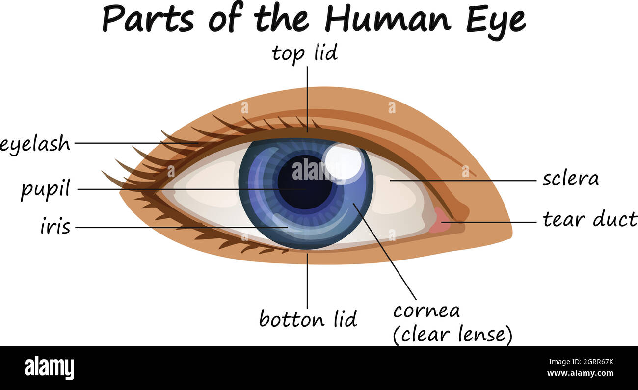
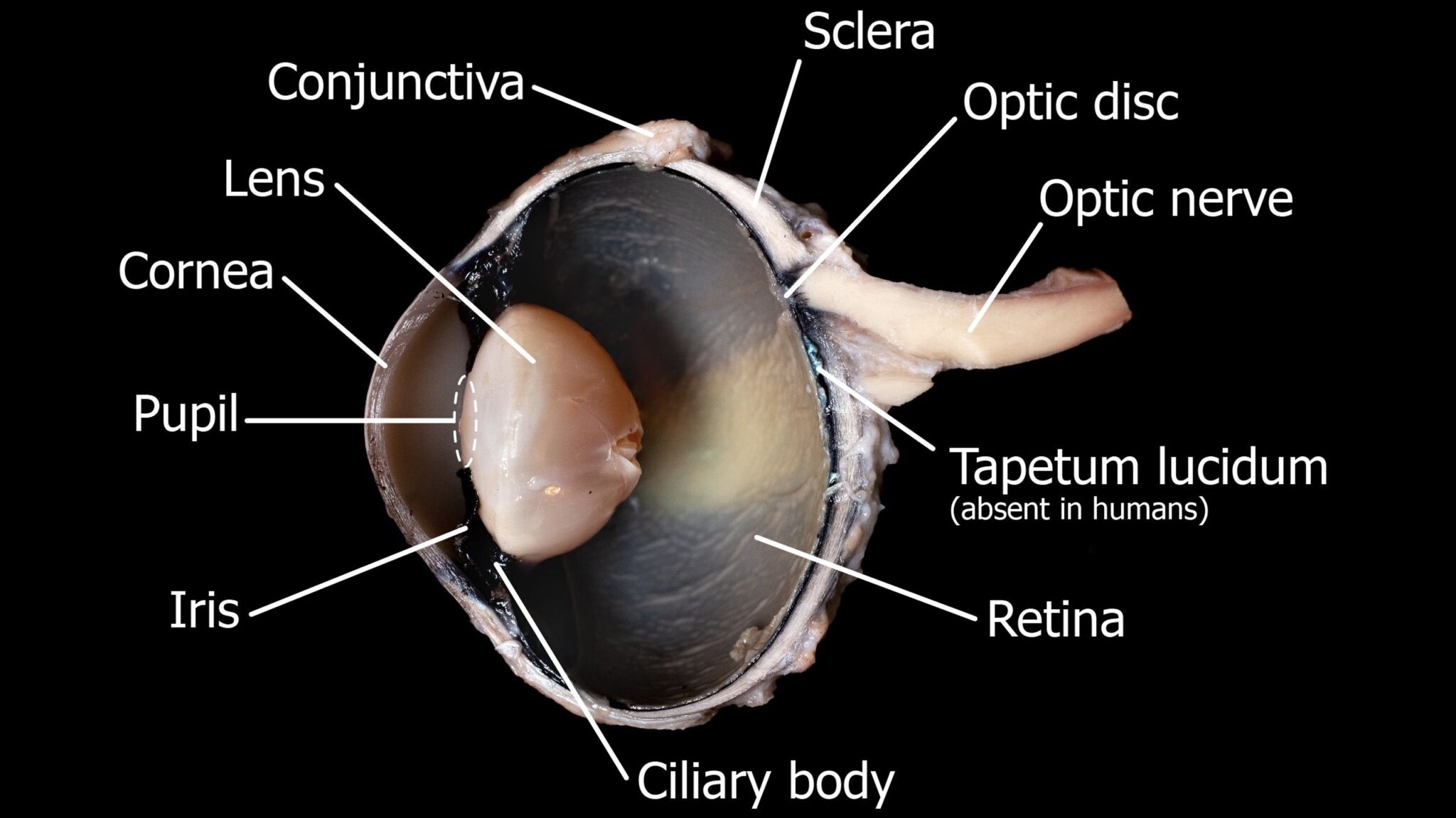
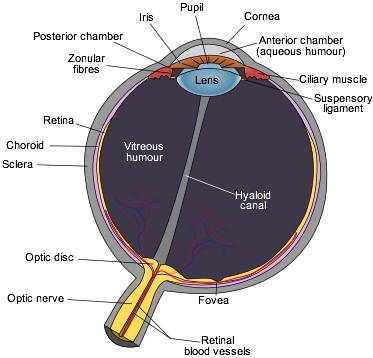
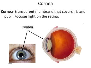
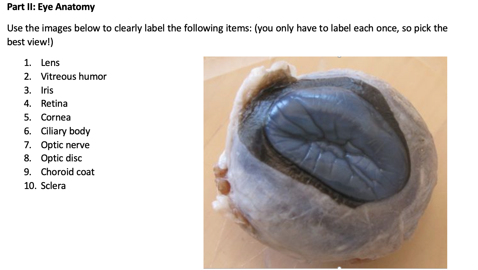
Cow's Eye Dissection - Eye diagram - Exploratorium A cow's iris is brown. Human irises come in many colors, including brown, blue, green, and gray. A clear fluid that helps the cornea keep its rounded shape. The pupil is the dark circle in the center of your iris. It's a hole that lets light into the inner eye. Your pupil is round. A cow's pupil is oval.
Eyes - Layers of Learning
Success Essays - Assisting students with assignments online Each paper writer passes a series of grammar and vocabulary tests before joining our team.
Eye Anatomy Activities Teaching Resources | Teachers Pay Teachers
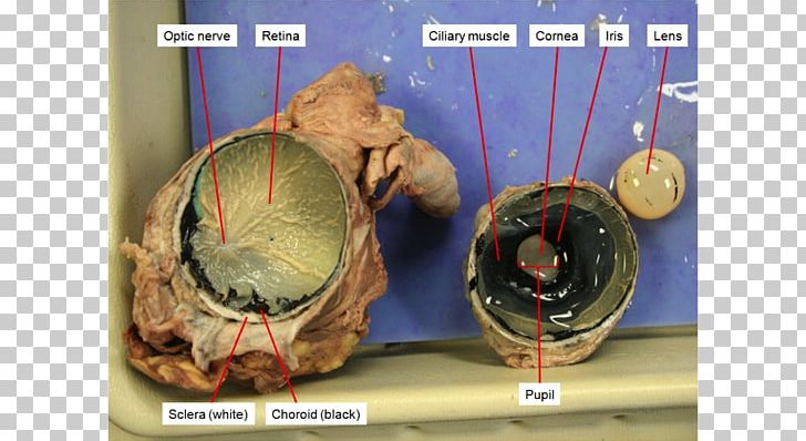
Parts and Functions - Dissecting A COW EYE Iris and Pupil. The iris is a muscle that controls how much light goes into the eye and suspended between the cornea and lens. As we have seen in the dissection a cow's iris is brown. The pupil is the dark circle that's in the center of the iris and it lets light into the inner eye.
Development, Anatomy and Physiology of the Eye The word ...
Cow Anatomy – External Body Parts and Internal Organs with Labeled Diagram Jul 28, 2021 · Cow anatomy labeled diagram. Here I would like to summarize the whole anatomical features of a cow (both internal and external) with the labeled diagram. I hope you will enjoy it and learn the anatomical features of the different organs of a cow. If you need more cow-labeled diagrams, you may join with anatomy learners on social media.
File:Eye (1).jpg - Wikimedia Commons
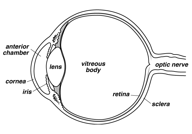
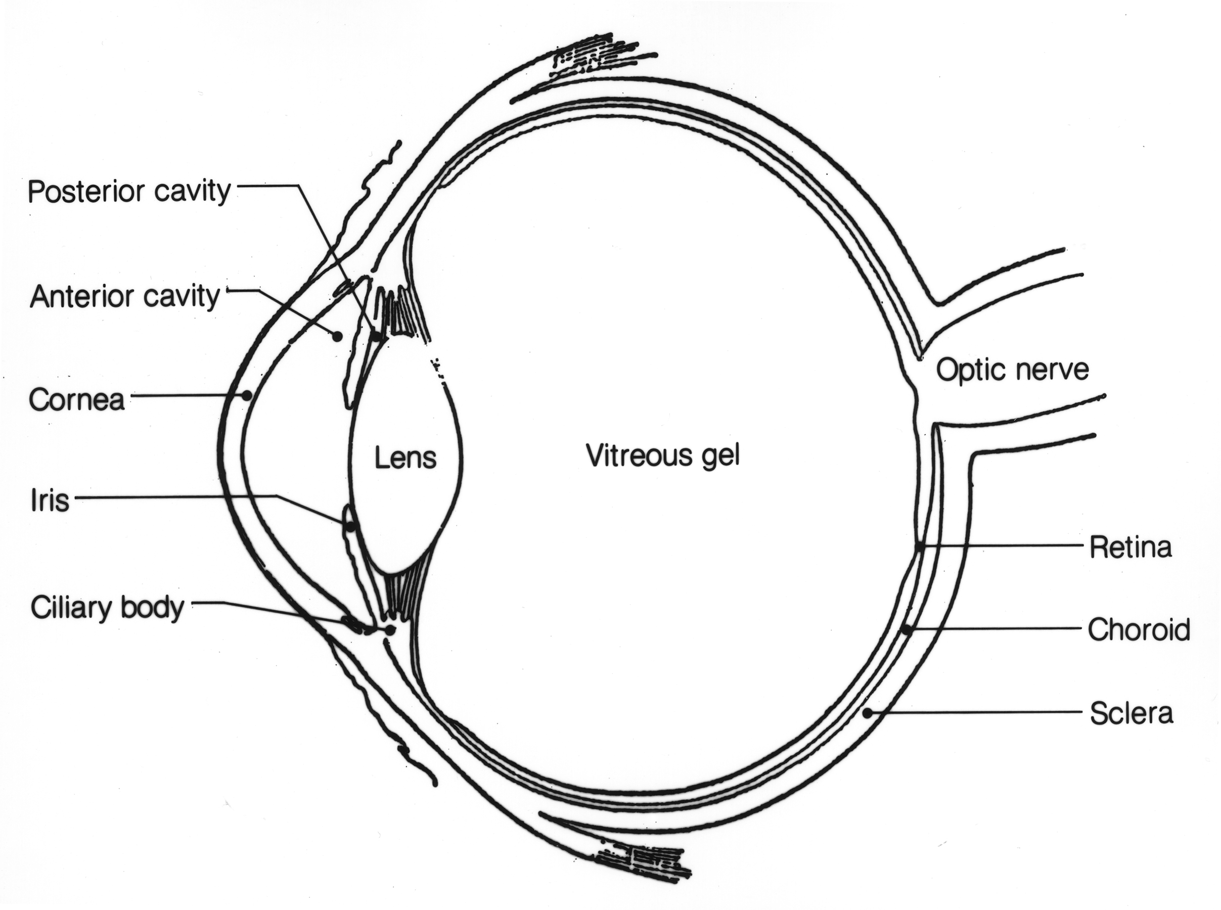
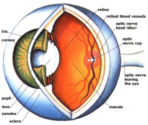
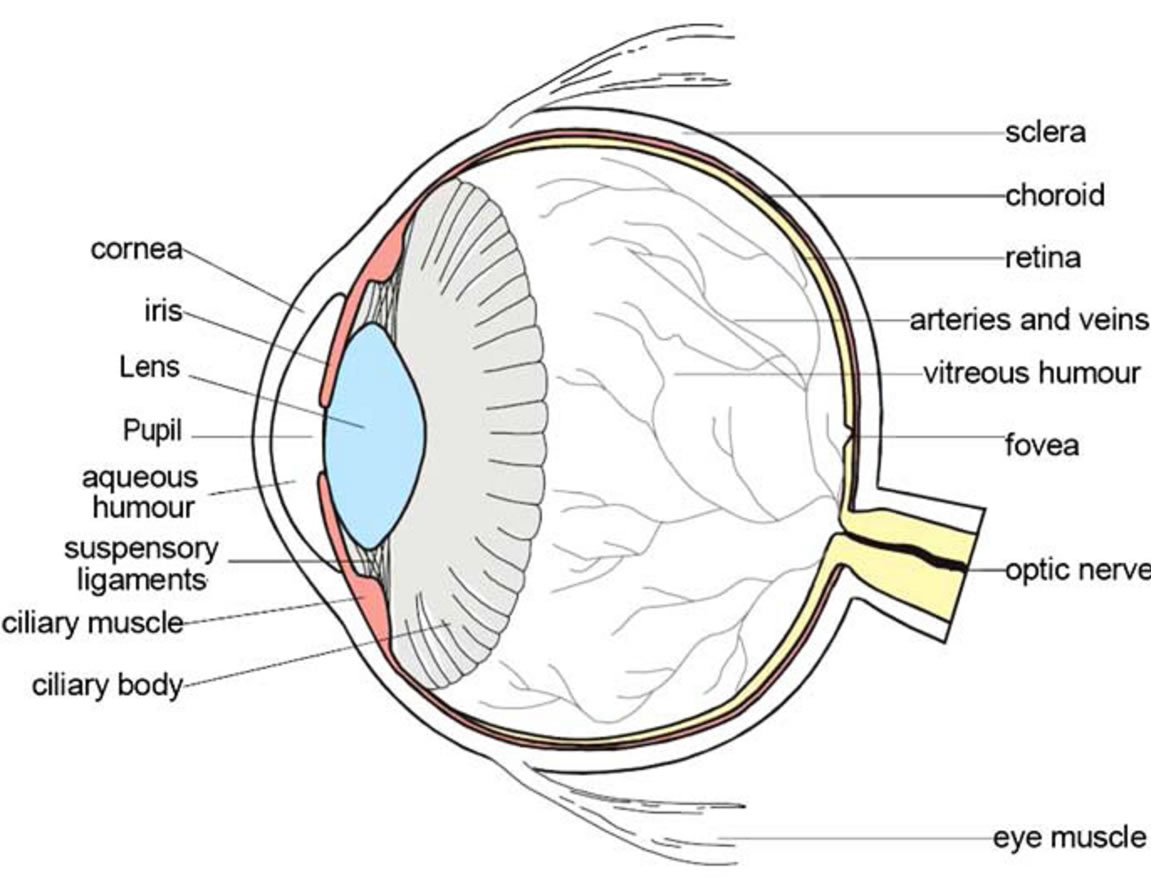
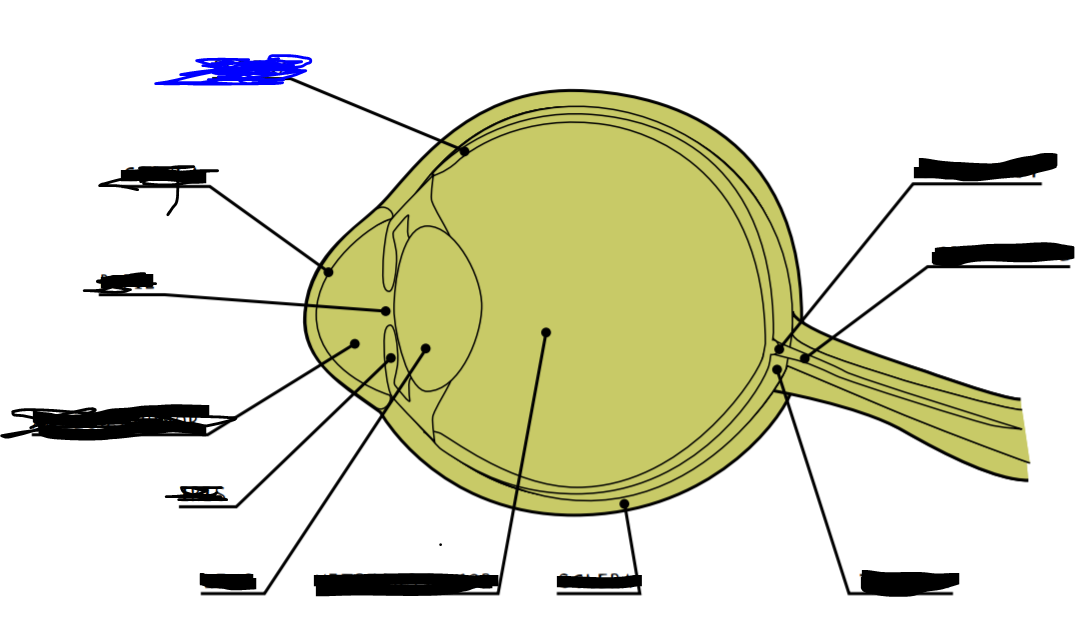
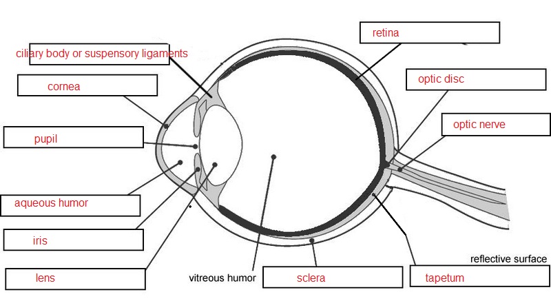
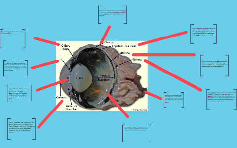
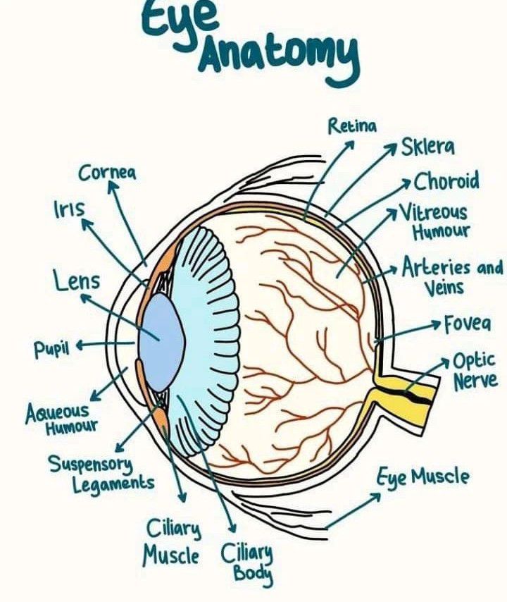
COW'S EYE dissection - Exploratorium This diagram shows the parts of the eye. Can you find these parts in a cow's eye? SCLERA TAPETUM OPTIC NERVE BLIND SPOT LENS VITREOUS HUMOR IRIS CORNEA RETINA AQUEOUS HUMOR PUPIL. COW'S EYE dissection page 3 ... The lens of the cow's eye feels soft on the outside and hard in the middle. Hold the lens up and look through it.
Body diagram label hi-res stock photography and images - Alamy
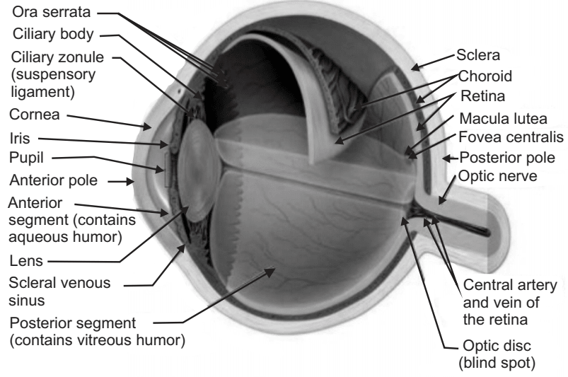
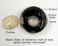
Cow Eye Dissection Guide - Google Slides Cow Eye. Use the point of a scissors or a scalpel to make an incision through the layers of the eye capsule (similar to figure 1); there are three layers from the exterior: sclera, whitish/grey, continuous with the transparent cornea, choroid, thin dark black layer and the retina, thin greyish/pink layer. Use a scissors to dissect the entire ...
Eye diagram parts hi-res stock photography and images - Alamy
Chuck Eye Steak Guide — What is it? How to Cook it? - Food … Mar 23, 2022 · The beef Chuck Eye steak comes from the chuck primal, which is the shoulder region.. The Ribeye is cut from the 6th to 12th rib of the cow, and the Chuck Eye is cut from the 5th. You’ll know from our beef cuts article that the shoulder and neck muscles are one of the hardest working parts of the cow, and as such, the muscles are generally tough.. The Chuck …

A&P Lab -- Unit 2 -- Labeled Cow Eye Diagram | Quizlet
Sheep Heart Dissection Lab for High School Science | HST Most heart diagrams show the left atrium and ventricle on the right side of the diagram. Imagine the heart in the body of a person facing you. The left side of their heart is on their left, but since you are facing them, it is on your right. 1. Identify the right and left sides of the heart.
Free Blank Eye Diagram, Download Free Blank Eye Diagram png ...
PDF Cow Eye Dissection: Examining Structure and Function - Woodstown During this activity, you will dissect a cow eye. You will observe several important features of the eye and develop your understanding of how each part functions to make vision possible. Materials ... Use the diagram to identify the internal structures of the eye. 6. Remove the vitreous humor and lens from the front portion of the eye. ...
SCB209 - Lab3 - Natural Sciences Open Educational Resources
Cow's Eye Dissection - Eye diagram - Exploratorium Learn how to dissect a cow's eye in your classroom. This resource includes: a step-by-step, hints and tips, a cow eye primer, and a glossary of terms. Cow's Eye Dissection - Eye diagram
Hamburg CSD - 5th Grade Cow Eyes
Cow Eye, Eye Histology, Eye Anatomy Diagram | Quizlet Start studying Cow Eye, Eye Histology, Eye Anatomy. Learn vocabulary, terms, and more with flashcards, games, and other study tools.

Anatomy of the Cow Eye DIAGRAM Diagram | Quizlet
Cow Anatomy - External Body Parts and Internal Organs with Labeled Diagram I will try to show you all of the body parts of a cow in labeled diagrams. The external body parts from the head region of a cow - in this head region, you might identify the mouth, lip, cheek, chin, muzzle, forehead, poll, ear, eye, nostril, and other.
Ocular Drug Delivery System - Solution Pharmacy
Cow Eye Dissection & Parts of the Eye Diagram | Quizlet Located in the back of the eye, contains the rods and cones. pupil The opening in the center of the iris through which light enters the eye lens The transparent structure behind the pupil that changes shape to help focus images on the retina. ligaments (eye) Fibers that connect ciliary muscles to the lens in the eye blind spot (optic disc)
Solved] 13. Label the Cow Eye (use your book or other ...
eye diagram labeled - anatomyhealth.z21.web.core.windows.net brain function structure functions diagram lobes macmillan labelled showing. Cow Eye Dissection - YouTube . cow eye dissection. Label The Muscles Of The Eye - PurposeGames . purposegames. 3d Eye Model 32 Pcs Assembled Human Anatomy Model New 3D Structure Of . auge. Photoreceptor Cell ...

10 Cow eye ideas | cow eyes, anatomy and physiology, eye anatomy
Using Genetic Algorithm for Identification of Diabetic ...
Parts of the eye | Quiz
Cow Eye Dissection
What is the purpose of eye diagrams? - Quora
Human eye diagram - Eye Anatomy - Online Biology Dictionary

COW EYE DISSECTION - NANDINI SONI

Parts and Functions - Dissecting A COW EYE
The Anatomy of the Cow Eye by Meli C
How the Eye Works | Western Laser Eye Associates

Interior Eye Anatomy - Interior Eye Anatomy Sclera Ciliary ...

Cow Eye Dissection & Anatomy Project | HST Learning Center
NEUR 320: Art and Vision
Sheep eye dissection
Cow Eye Dissection & Anatomy Project | HST Learning Center

What are the parts of the eye? - ppt download
Solved Please label each diagram with the appropriate | Chegg.com
Eye anatomy - MEDizzy
Eye Structure and Function in Cats - Cat Owners - MSD ...

Cow Eye Dissection | Carolina.com
Anatomy_of_the Eye
Post a Comment for "39 cow eye diagram labeled"